File:Confocal microscopy of midbrain vasculature and neural tissue of a 3-dpf zebrafish larva - journal.pbio.1001374.s019.mov.ogv
Jump to navigation
Jump to search
Size of this JPG preview of this OGG file: 800 × 586 pixels. Other resolutions: 320 × 234 pixels | 640 × 469 pixels | 1,024 × 750 pixels.
Original file (Ogg Theora video file, length 17 s, 1,024 × 750 pixels, 680 kbps, file size: 1.36 MB)
Captions
Captions
Add a one-line explanation of what this file represents
Summary
[edit]| Description |
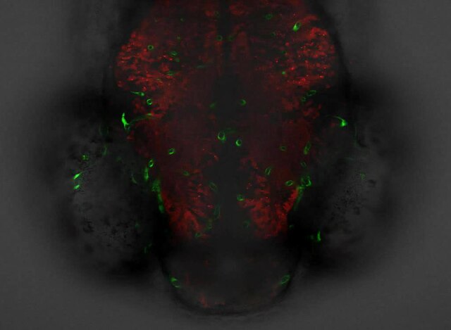
English: A z -stack movie obtained by a confocal microscope showing both the midbrain vasculature (green) and neural tissue (red) of a 3-dpf Tg(kdrl:eGFP,HuC:gal4-uas-mCherry) zebrafish larva. At the beginning of the movie, its projected image, which is shown in the bottom panel of Figure 1A , is presented. The direction of the movie is from ventral to dorsal. Dorsal view, anterior is down.
(MOV) |
||
| Date | |||
| Source | Video S1 from Chen Q, Jiang L, Li C, Hu D, Bu J, Cai D, Du J, Krasnow M. "Haemodynamics-Driven Developmental Pruning of Brain Vasculature in Zebrafish". PLoS Biology. DOI:10.1371/journal.pbio.1001374. PMID 22904685. PMC: 3419171. | ||
| Author | Chen Q, Jiang L, Li C, Hu D, Bu J, Cai D, Du J, Krasnow M | ||
| Permission (Reusing this file) |
This file is licensed under the Creative Commons Attribution 3.0 Unported license.
|
||
| Provenance |
|
File history
Click on a date/time to view the file as it appeared at that time.
| Date/Time | Thumbnail | Dimensions | User | Comment | |
|---|---|---|---|---|---|
| current | 22:56, 21 August 2012 | 17 s, 1,024 × 750 (1.36 MB) | Daniel Mietchen (talk | contribs) | Uploaded with the Open Access Media Importer. (test edit) botrequest |
You cannot overwrite this file.
File usage on Commons
The following page uses this file:
- File:Pbio.1001374.s019.mov.ogv (file redirect)