File:Disporella guada (10.5852-ejt.2021.773.1507) Figure 2.png
Jump to navigation
Jump to search

Size of this preview: 525 × 599 pixels. Other resolutions: 210 × 240 pixels | 421 × 480 pixels | 673 × 768 pixels | 897 × 1,024 pixels | 1,892 × 2,159 pixels.
Original file (1,892 × 2,159 pixels, file size: 3.01 MB, MIME type: image/png)
Captions
Captions
Add a one-line explanation of what this file represents
Summary
[edit]_Figure_2.png&action=edit§ion=1)
| Description |
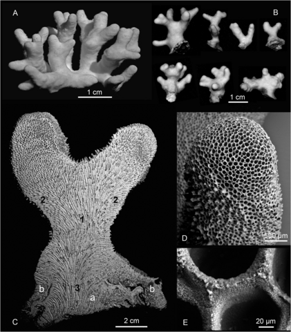
English: Fig. 2. Photographs (A–B) and scanning electron micrographs of Disporella guada Harmelin, Taylor & Waeschenbach sp. nov. A. Holotype, specimen kept dry (MNHN-IB-2017-696). B. Seven variously shaped colonies (top row, left to right: NHMUK 2021.2.25.1, 2021.3.19.2, 2018.1.15.63, 2021.3.19.1; bottom row, left to right: NHMUK 2021.6.14.1, 2021.6.14.2, 2021.6.14.3). C. Longitudinal section of a specimen (NHMUK 2021.3.19.1) with two branches; 1: endozone, 2: exozone, 3: basal part with central primary attachment zone (3a) and secondary peripheral attachment zone (3b). D–E. NHMUK 2021.3.19.2. D. Growing tip. E. Interzooecial walls at growing tip. |
| Date | |
| Source | https://doi.org/10.5852/ejt.2021.773.1507 |
| Author | Taylor, P. D., Harmelin, J.-G., Waeschenbach, A., & Bouchon, C. (2021). Disporella guada sp. nov., an erect-ramose rectangulate cyclostome (Bryozoa, Stenolaemata) from the Caribbean Sea: convergent evolution in bryozoan colony morphology. European Journal of Taxonomy, 773(1), 1–18. |
| Permission (Reusing this file) |
This file is licensed under the Creative Commons Attribution 4.0 International license.
|
File history
Click on a date/time to view the file as it appeared at that time.
| Date/Time | Thumbnail | Dimensions | User | Comment | |
|---|---|---|---|---|---|
| current | 19:47, 20 February 2022 | | 1,892 × 2,159 (3.01 MB) | Christian Ferrer (talk | contribs) | {{Information | description = {{en|1=Fig. 2. Photographs (A–B) and scanning electron micrographs of ''Disporella guada'' Harmelin, Taylor & Waeschenbach sp. nov. A. Holotype, specimen kept dry (MNHN-IB-2017-696). B. Seven variously shaped colonies (top row, left to right: NHMUK 2021.2.25.1, 2021.3.19.2, 2018.1.15.63, 2021.3.19.1; bottom row, left to right: NHMUK 2021.6.14.1, 2021.6.14.2, 2021.6.14.3). C. Longitudinal section of a specimen (NHMUK 2021.3.19.1) with two branches; 1: endozone, 2... |
You cannot overwrite this file.
File usage on Commons
There are no pages that use this file.
_Figure_2.png&oldid=631242019)