File:Embryonic-Medaka-Model-of-Microglia-in-the-Developing-CNS-Allowing-In-Vivo-Analysis-of-Their-pone.0127325.s010.ogv
Jump to navigation
Jump to search
Size of this JPG preview of this OGG file: 800 × 440 pixels. Other resolutions: 320 × 176 pixels | 640 × 352 pixels | 872 × 480 pixels.
Original file (Ogg multiplexed audio/video file, Theora/Vorbis, length 15 s, 872 × 480 pixels, 851 kbps overall, file size: 1.51 MB)
Captions
Captions
Add a one-line explanation of what this file represents
Summary
[edit]| Description |
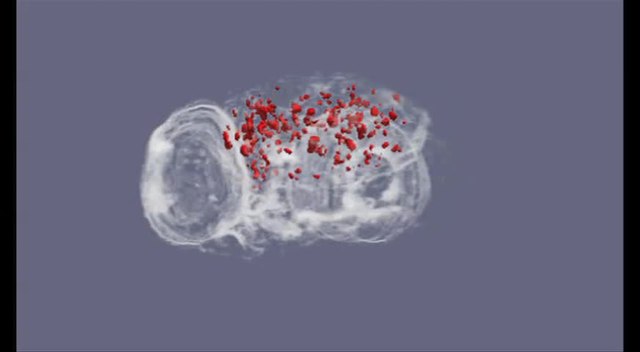
English: 3D images of ApoE in an irradiated p53 -/- embryo 24 h after irradiation. This video shows 3D images of ApoE in the brain (excluding the eyes) of an irradiated p53-/- embryo 24 h after irradiation (red dots). |
||
| Date | |||
| Source | S4 Movie from Yasuda T, Oda S, Hibi Y, Satoh S, Nagata K, Hirakawa K, Kutsuna N, Sagara H, Mitani H (2015). "Embryonic Medaka Model of Microglia in the Developing CNS Allowing In Vivo Analysis of Their Spatiotemporal Recruitment in Response to Irradiation". PLOS ONE. DOI:10.1371/journal.pone.0127325. PMID 26061282. PMC: 4465025. | ||
| Author | Yasuda T, Oda S, Hibi Y, Satoh S, Nagata K, Hirakawa K, Kutsuna N, Sagara H, Mitani H | ||
| Permission (Reusing this file) |
This file is licensed under the Creative Commons Attribution 4.0 International license.
|
||
| Provenance |
|
File history
Click on a date/time to view the file as it appeared at that time.
| Date/Time | Thumbnail | Dimensions | User | Comment | |
|---|---|---|---|---|---|
| current | 03:52, 28 June 2015 | 15 s, 872 × 480 (1.51 MB) | Open Access Media Importer Bot (talk | contribs) | Automatically uploaded media file from Open Access source. Please report problems or suggestions here. |
You cannot overwrite this file.
File usage on Commons
The following 2 pages use this file: