File:Experimental-Cerebral-Malaria-Pathogenesis—Hemodynamics-at-the-Blood-Brain-Barrier-ppat.1004528.s042.ogv
Jump to navigation
Jump to search
Size of this JPG preview of this OGG file: 597 × 599 pixels. Other resolutions: 239 × 240 pixels | 478 × 480 pixels | 816 × 819 pixels.
Original file (Ogg Theora video file, length 6.1 s, 816 × 819 pixels, 1.91 Mbps, file size: 1.38 MB)
Captions
Captions
Add a one-line explanation of what this file represents
Summary
[edit]| Description |
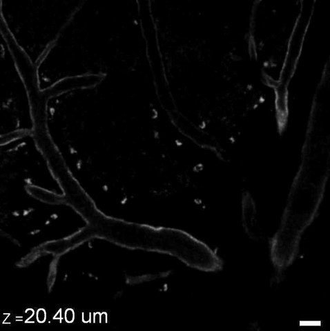
English: ICAM-1 expression in an uninfected control mouse. Intravital microscopy stack showing a lower level of ICAM-1 (gray) expression on the surface of postcapillary venules compared to mice with ECM (Video S12) or hyperparasitemia (Video S13). ICAM-1 expressing leukocytes are absent. Scale bar = 20 µm. |
||
| Date | |||
| Source | Video S19 from Nacer A, Movila A, Sohet F, Girgis N, Gundra U, Loke P, Daneman R, Frevert U (2014). "Experimental Cerebral Malaria Pathogenesis—Hemodynamics at the Blood Brain Barrier". PLOS Pathogens. DOI:10.1371/journal.ppat.1004528. PMID 25474413. PMC: 4256476. | ||
| Author | Nacer A, Movila A, Sohet F, Girgis N, Gundra U, Loke P, Daneman R, Frevert U | ||
| Permission (Reusing this file) |
This file is licensed under the Creative Commons Attribution 4.0 International license.
|
||
| Provenance |
|
File history
Click on a date/time to view the file as it appeared at that time.
| Date/Time | Thumbnail | Dimensions | User | Comment | |
|---|---|---|---|---|---|
| current | 22:55, 13 December 2014 | 6.1 s, 816 × 819 (1.38 MB) | Open Access Media Importer Bot (talk | contribs) | Automatically uploaded media file from Open Access source. Please report problems or suggestions here. |
You cannot overwrite this file.
File usage on Commons
There are no pages that use this file.