File:Gray727.svg

Sākotnējais fails (SVG fails, definētais izmērs 1 025 × 598 pikseļi, faila izmērs: 18 KB)
Captions
Captions
Kopsavilkums
[labot šo sadaļu]| Apraksts |
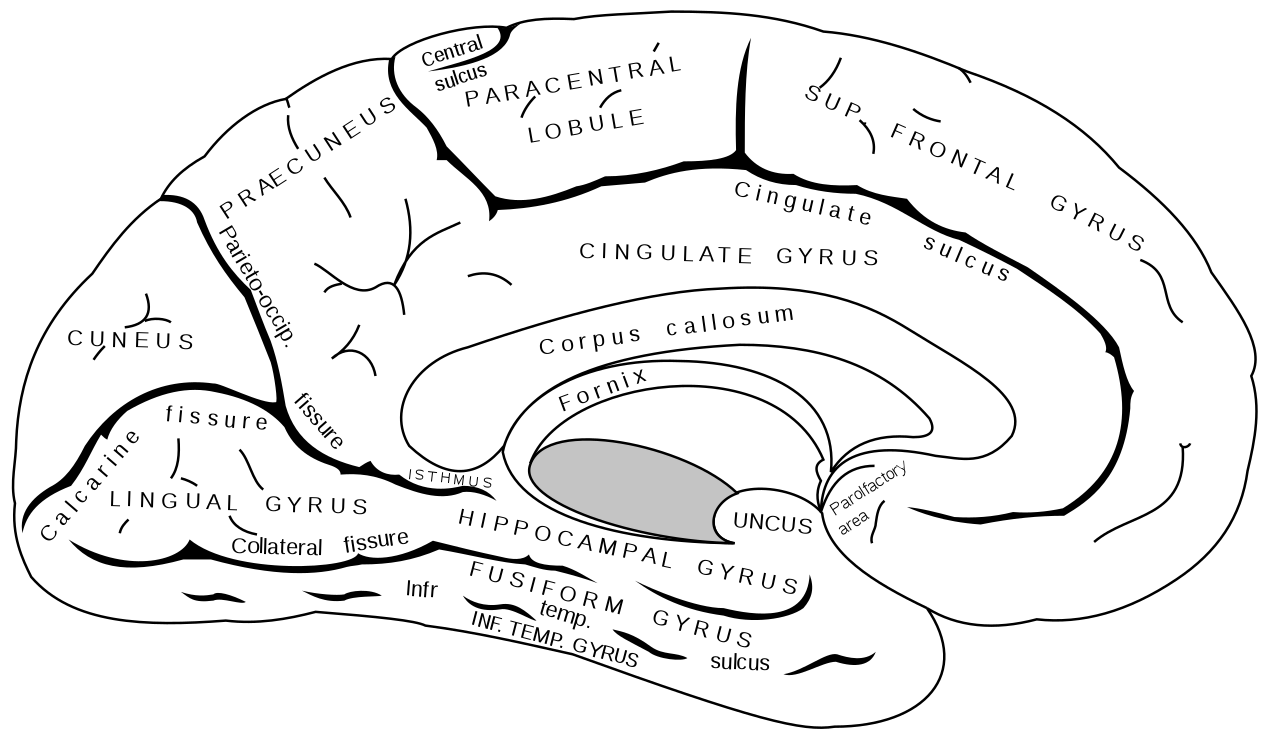
English: Medial surface of left cerebral hemisphere. |
||||||||||||||||||||
| Plate | 727 | ||||||||||||||||||||
| Datums | before 1858. gada | ||||||||||||||||||||
| Avots |
|
||||||||||||||||||||
| Autors |
|
||||||||||||||||||||
| Citas versijas |
Derivative works of this file: |
||||||||||||||||||||
.jpg)

|
This SVG file contains embedded text that can be translated into your language, using any capable SVG editor, text editor or the SVG Translate tool. For more information see: About translating SVG files. |
Grāmata
[labot šo sadaļu]| Henry Gray: Greja Anatomija, 20. izdevums
|
|||||||||||||||||||||||
|---|---|---|---|---|---|---|---|---|---|---|---|---|---|---|---|---|---|---|---|---|---|---|---|
| Autors |
|
-_Title_page.png) | |||||||||||||||||||||
| Redaktors |
Revised by Warren H. Lewis |
||||||||||||||||||||||
| Ilustrators |
|
||||||||||||||||||||||
| Nosaukums | |||||||||||||||||||||||
| Izdevums |
20 |
||||||||||||||||||||||
| Izdevējs | |||||||||||||||||||||||
| Object type |
versija, izdevums vai tulkojums |
||||||||||||||||||||||
| Page overview | list of all the plates | ||||||||||||||||||||||
| Valoda |
angļu valoda |
||||||||||||||||||||||
| Publicēšanas datums |
1918. gada |
||||||||||||||||||||||
| Izdošanas vieta |
Filadelfija / Ņujorka |
||||||||||||||||||||||
| Avots | Bartleby | ||||||||||||||||||||||

Licence
[labot šo sadaļu]
This image is in the public domain because it is a mere mechanical scan or photocopy of a public domain original, or – from the available evidence – is so similar to such a scan or photocopy that no copyright protection can be expected to arise. The original itself is in the public domain for the following reason:
This tag is designed for use where there may be a need to assert that any enhancements (eg brightness, contrast, colour-matching, sharpening) are in themselves insufficiently creative to generate a new copyright. It can be used where it is unknown whether any enhancements have been made, as well as when the enhancements are clear but insufficient. For known raw unenhanced scans you can use an appropriate {{PD-old}} tag instead. For usage, see Commons:When to use the PD-scan tag.  | ||||
Faila hronoloģija
Uzklikšķini uz datums/laiks kolonnā esošās saites, lai apskatītos, kā šis fails izskatījās tad.
| Datums/Laiks | Attēls | Izmēri | Dalībnieks | Komentārs | |
|---|---|---|---|---|---|
| tagadējais | 2009. gada 25. oktobris, plkst. 10.41 | | 1 025 × 598 (18 KB) | Was a bee (diskusija | devums) | +text (collateral fissure) |
| 2006. gada 30. decembris, plkst. 18.20 |  | 1 025 × 598 (11 KB) | Mysid (diskusija | devums) | typo (gurys -> gyrus) | |
| 2006. gada 29. novembris, plkst. 08.27 |  | 1 025 × 598 (11 KB) | Mysid (diskusija | devums) | oops. let's render first. | |
| 2006. gada 29. novembris, plkst. 08.26 |  | 1 025 × 598 (11 KB) | Mysid (diskusija | devums) | repairing spaces | |
| 2006. gada 29. novembris, plkst. 08.21 |  | 1 025 × 598 (11 KB) | Mysid (diskusija | devums) | ==Summary== {{Information |Description=Medial surface of left cerebral hemisphere. Figure 727 from Gray's Anatomy. |Source=Vectorized in CorelDraw by Mysid, based on the online version of the 1918 Gray's Anatomy. |Date=November 29, 2006 | |
Šo failu nevar pārrakstīt.
Faila lietojums
Šo failu izmanto šajās 36 lapās:
- Gyri
- Sulci (neuroanatomy)
- File:Gray726.svg
- File:Gray727.png
- File:Gray727.svg
- File:Gray727 Cuneus.png
- File:Gray727 anterior cingulate cortex.png
- File:Gray727 calcarine sulcus.svg
- File:Gray727 cant been seen medial.png
- File:Gray727 central sulcus.svg
- File:Gray727 cingulate gyrus.png
- File:Gray727 cingulate sulcus.svg
- File:Gray727 collateral fissure.svg
- File:Gray727 frontal pole.png
- File:Gray727 fusiform gyrus.png
- File:Gray727 inferior temporal gyrus.png
- File:Gray727 inferior temporal sulcus.svg
- File:Gray727 lateral fissure.svg
- File:Gray727 latin.svg
- File:Gray727 lingual gyrus.png
- File:Gray727 marginal sulcus.svg
- File:Gray727 occipital lobe.png
- File:Gray727 occipital pole.png
- File:Gray727 paracentral gyrus.png
- File:Gray727 paracentral sulcus.png
- File:Gray727 parahippocampal gyrus.png
- File:Gray727 parieto-occipital fissure.svg
- File:Gray727 precuneus.png
- File:Gray727 subparietal sulcus.svg
- File:Gray727 sulcus of the corpus callosum.svg
- File:Gray727 superior frontal gyrus.png
- File:Gray727 temporal pole.png
- File:Gray727 uncus of parahippocampal gyrus.png
- File:Gyrus cinguli.png
- File:Sulcus centralis - Identification axial - MRI T2.jpg
- File:Sulcus centralis - Identification sagittal - MRI T2.jpg
Globālais faila lietojums
Šīs Vikipēdijas izmanto šo failu:
- Izmantojums ar.wikipedia.org
- Izmantojums ast.wikipedia.org
- Izmantojums cs.wikipedia.org
- Izmantojums de.wikipedia.org
- Izmantojums en.wikipedia.org
- Izmantojums es.wikipedia.org
- Izmantojums et.wikipedia.org
- Izmantojums hr.wikipedia.org
- Izmantojums id.wikipedia.org
- Izmantojums it.wikipedia.org
- Izmantojums ja.wikipedia.org
- Izmantojums kk.wikipedia.org
- Izmantojums nl.wikipedia.org
- Izmantojums pl.wikipedia.org
- Izmantojums pt.wikipedia.org
- Izmantojums ru.wikipedia.org
- Izmantojums uk.wikipedia.org
- Izmantojums zh.wikipedia.org