File:Histology of paneth cells, annotated.jpg
Une page de Wikimedia Commons, la médiathèque libre.
Aller à la navigation
Aller à la recherche
- Fichier
- Historique du fichier
- Utilisations locales du fichier
- Utilisations du fichier sur d’autres wikis
- Métadonnées

Taille de cet aperçu : 675 × 600 pixels. Autres résolutions : 270 × 240 pixels | 540 × 480 pixels | 864 × 768 pixels | 1 257 × 1 117 pixels.
Fichier d’origine (1 257 × 1 117 pixels, taille du fichier : 367 kio, type MIME : image/jpeg)
Informations sur le fichier
Données structurées
Légendes
Légendes
Ajoutez en une ligne la description de ce que représente ce fichier
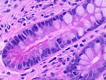
Histology of paneth cells, located at the base of the crypts of the small intestinal mucosa, and displaying bright red cytoplasmic granules. H&E stain.
Description
[modifier]| DescriptionHistology of paneth cells, annotated.jpg |
English: Histology of Paneth cells, located at the base of the crypts of the small intestinal mucosa, and displaying merocrine secretion of bright red cytoplasmic granules. H&E stain. - Source for merocrine: Matsubara F (1977). "Morphological study of the Paneth cell. Paneth cells in intestinal metaplasia of the stomach and duodenum of man.". Acta Pathol Jpn 27 (5): 677-95. DOI:10.1111/j.1440-1827.1977.tb00185.x. PMID 930588. |
| Date | |
| Source | Travail personnel |
| Auteur |
 - Reusing images - Conflicts of interest: None Consent note: Consent from the patient or patient's relatives is regarded as redundant, because of absence of identifiable features (List of HIPAA identifiers) in the media and case information (See also HIPAA case reports guidance). |
| Autres versions |
 |
Conditions d’utilisation
[modifier]| Ce fichier est disponible selon les termes de la licence Creative Commons CC0 Don universel au domaine public. | |
| La personne qui a associé une œuvre avec cet acte l’a placée dans le domaine public en renonçant mondialement à tous ses droits sur cette œuvre en vertu des lois relatives au droit d’auteur, ainsi qu’à tous les droits juridiques connexes et voisins qu’elle possédait sur l’œuvre, sans autre limite que celles imposées par la loi. Vous pouvez copier, modifier, distribuer et utiliser cette œuvre, y compris à des fins commerciales, sans qu’il soit nécessaire d’en demander la permission.
http://creativecommons.org/publicdomain/zero/1.0/deed.enCC0Creative Commons Zero, Public Domain Dedicationfalsefalse |
Historique du fichier
Cliquer sur une date et heure pour voir le fichier tel qu'il était à ce moment-là.
| Date et heure | Vignette | Dimensions | Utilisateur | Commentaire | |
|---|---|---|---|---|---|
| actuel | 23 novembre 2020 à 18:13 |  | 1 257 × 1 117 (367 kio) | Mikael Häggström (d | contributions) | Uploaded a work by {{Mikael Häggström|cat=Micrographs of the gastrointestinal tract|consent=noid}}}} from {{Own}} with UploadWizard |
Vous ne pouvez pas remplacer ce fichier.
Utilisations locales du fichier
La page suivante utilise ce fichier :
Utilisations du fichier sur d’autres wikis
Les autres wikis suivants utilisent ce fichier :
- Utilisation sur en.wikipedia.org
- Utilisation sur www.wikidata.org
Métadonnées
Ce fichier contient des informations supplémentaires, probablement ajoutées par l'appareil photo numérique ou le numériseur utilisé pour le créer.
Si le fichier a été modifié depuis son état original, certains détails peuvent ne pas refléter entièrement l'image modifiée.
| Fabricant de l’appareil photo | Olympus |
|---|---|
| Modèle de l’appareil photo | LC30 |
| Auteur | 623546 |
| Durée d’exposition | 7 424/116 447 s (0,063754326002387 s) |
| Date et heure de génération des données | 23 novembre 2020 à 13:04 |
| Orientation | Normale |
| Résolution horizontale | 72 pt/po |
| Résolution verticale | 72 pt/po |
| Date de modification du fichier | 23 novembre 2020 à 13:04 |
| Version d’EXIF | 2.1 |
| Date et heure de la numérisation | 23 novembre 2020 à 13:04 |
| Ouverture APEX | 0,1 |
| Distance du sujet | 0,0185 mètre |
| Mode de mesure | Moyenne |
| Fractions de secondes de l’horodatage de la prise de vue originale | 00 |
| Fractions de secondes de l’horodatage de la numérisation | 00 |
| Version de FlashPix prise en charge | 1 |
| Espace colorimétrique | sRGB |
| Mode d’exposition | Exposition manuelle |