File:Image from page 235 of "On the anatomy of vertebrates (electronic resource)" (1866) (14755203772).jpg
Original file (860 × 2,296 pixels, file size: 449 KB, MIME type: image/jpeg)
Captions
Captions
Summary[edit]
%22_(1866)_(14755203772).jpg&action=edit§ion=1)
| Description |
Identifier: b20416039_001 Title: On the anatomy of vertebrates [electronic resource] Year: 1866 (1860s) Authors: Owen, Richard, 1804-1892 Subjects: Anatomy, Comparative Vertebrates Fishes Reptiles Mammals Birds Publisher: London : Longmans, Green Contributing Library: Wellcome Library Digitizing Sponsor: Wellcome Library
Click here to view book online to see this illustration in context in a browseable online version of this book.
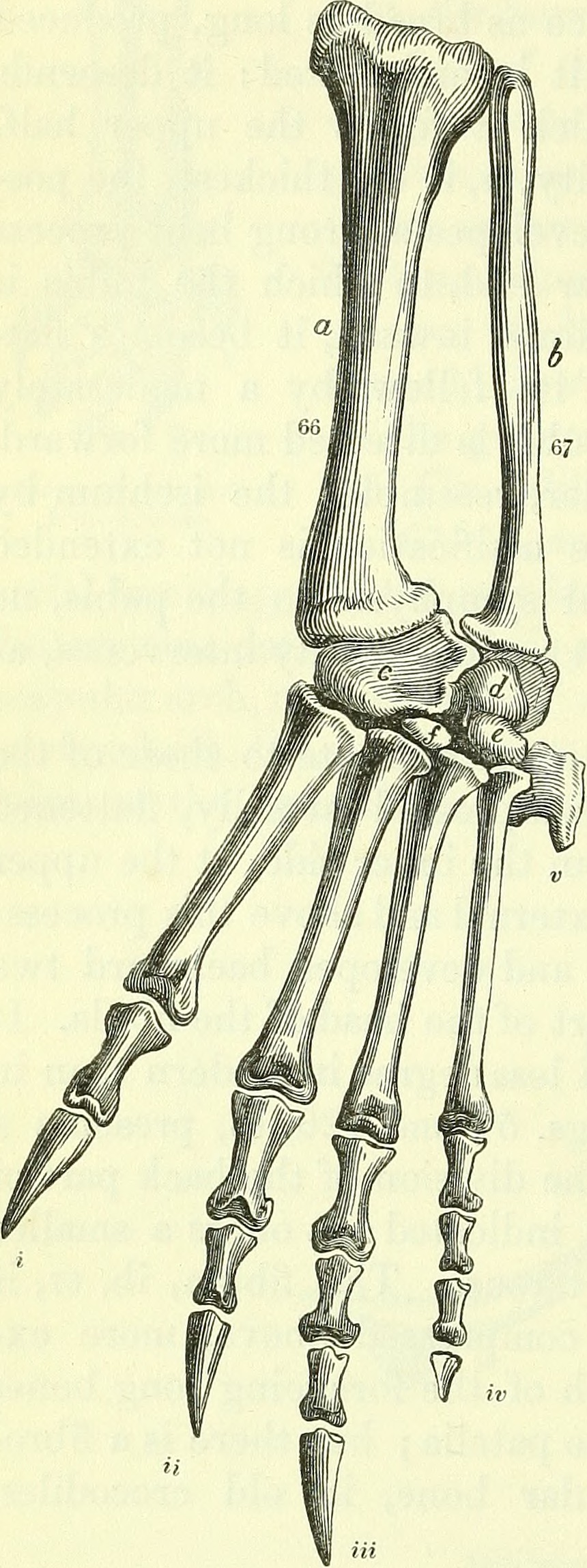
Text Appearing After Image: Bones of leg and foot, Crocodile ANATOMY OF VERTEBRATES. 191
|
| Date | |
| Source | Image from page 235 of "On the anatomy of vertebrates [electronic resource]" (1866) |
| Author | Internet Archive Book Images |
| Permission (Reusing this file) |
Internet Archive Book Images @ Flickr Commons |
Licensing[edit]
%22_(1866)_(14755203772).jpg&action=edit§ion=2)
This image was taken from Flickr's The Commons. The uploading organization may have various reasons for determining that no known copyright restrictions exist, such as:
More information can be found at https://flickr.com/commons/usage/. Please add additional copyright tags to this image if more specific information about copyright status can be determined. See Commons:Licensing for more information. |
| This image was originally posted to Flickr by Internet Archive Book Images at https://flickr.com/photos/126377022@N07/14755203772. It was reviewed on 7 June 2017 by FlickreviewR and was confirmed to be licensed under the terms of the No known copyright restrictions. |
File history
Click on a date/time to view the file as it appeared at that time.
| Date/Time | Thumbnail | Dimensions | User | Comment | |
|---|---|---|---|---|---|
| current | 17:08, 7 June 2017 | 860 × 2,296 (449 KB) | Jarble (talk | contribs) | Transferred from Flickr via Flickr2Commons |
You cannot overwrite this file.
File usage on Commons
There are no pages that use this file.
%22_(1866)_(14755203772).jpg&oldid=472560186)