File:Imaging-of-human-differentiated-3D-neural-aggregates-using-light-sheet-fluorescence-microscopy-Movie3.ogv
Jump to navigation
Jump to search
Size of this JPG preview of this OGG file: 727 × 600 pixels. Other resolutions: 291 × 240 pixels | 582 × 480 pixels | 931 × 768 pixels | 1,068 × 881 pixels.
Original file (Ogg Theora video file, length 5.0 s, 1,068 × 881 pixels, 2.3 Mbps, file size: 1.37 MB)
Captions
Captions
Add a one-line explanation of what this file represents
Summary
[edit]| Description |
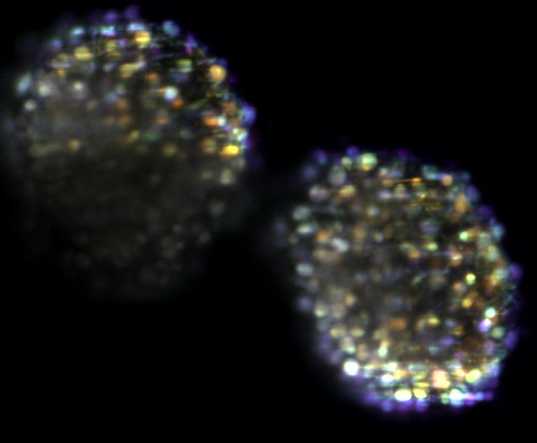
English: Tracking of the Ca2+ oscillations in two neural aggregates stained with AM-ester calcium indicator Fluo-4 over 25 min every 6 s. Here is represented the maximum intensity projection of five different planes 2 μm apart, plotted with different color. |
||
| Date | |||
| Source | Movie S3 from Gualda E, Simao D, Pinto C, Alves P, Brito C (2014). "Imaging of human differentiated 3D neural aggregates using light sheet fluorescence microscopy". Frontiers in Cellular Neuroscience. DOI:10.3389/fncel.2014.00221. PMID 25161607. PMC: 4123789. | ||
| Author | Gualda E, Simao D, Pinto C, Alves P, Brito C | ||
| Permission (Reusing this file) |
This file is licensed under the Creative Commons Attribution 3.0 Unported license.
|
||
| Provenance |
|
File history
Click on a date/time to view the file as it appeared at that time.
| Date/Time | Thumbnail | Dimensions | User | Comment | |
|---|---|---|---|---|---|
| current | 23:36, 28 August 2014 | 5.0 s, 1,068 × 881 (1.37 MB) | Open Access Media Importer Bot (talk | contribs) | Automatically uploaded media file from Open Access source. Please report problems or suggestions here. |
You cannot overwrite this file.
File usage on Commons
There are no pages that use this file.