File:Loupe-binoculaire-p1030891.jpg
跳至導覽
跳至搜尋
預覽大小:800 × 594 像素。 其他解析度:320 × 238 像素 | 640 × 475 像素 | 1,024 × 760 像素 | 1,280 × 950 像素 | 2,535 × 1,882 像素。
原始檔案 (2,535 × 1,882 像素,檔案大小:2.72 MB,MIME 類型:image/jpeg)
說明
說明
添加單行說明來描述出檔案所代表的內容
摘要[編輯]
| 描述 |
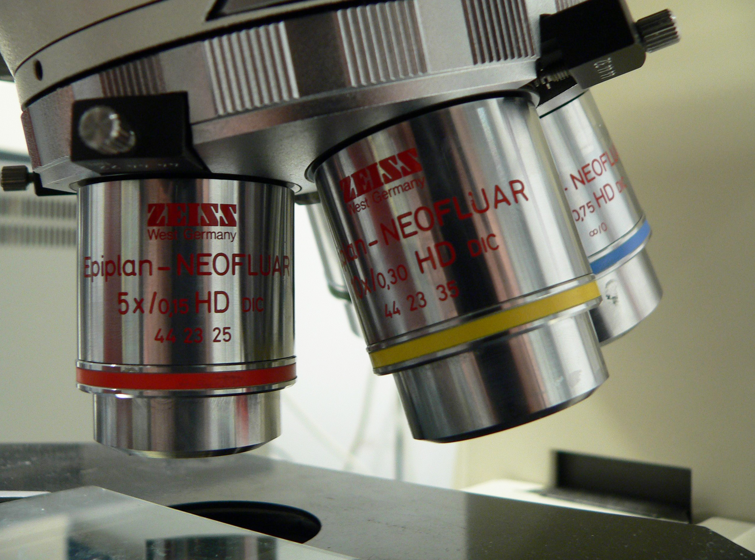
English: binocular microscope
Français : Loupe binoculaire |
||
| 日期 | 日期不明 | ||
| 來源 | 自己的作品 | ||
| 作者 | Rama | ||
| 授權許可 (重用此檔案) |
此檔案採用創用CC 姓名標示-相同方式分享 2.0 法國授權條款。
 |

檔案歷史
點選日期/時間以檢視該時間的檔案版本。
| 日期/時間 | 縮圖 | 尺寸 | 用戶 | 備註 | |
|---|---|---|---|---|---|
| 目前 | 2015年12月19日 (六) 15:13 | | 2,535 × 1,882(2.72 MB) | Jacek Halicki(對話 | 貢獻) | tilt, more light, jpg compression |
| 2006年3月17日 (五) 13:43 |  | 2,560 × 1,920(546 KB) | Rama(對話 | 貢獻) | {{fr|Loupe binoculaire}} {{en|binocular microscope}} {{Rama}} Category:Microscopes |
無法覆蓋此檔案。
檔案用途
下列2個頁面有用到此檔案:
全域檔案使用狀況
以下其他 wiki 使用了這個檔案:
- af.wikipedia.org 的使用狀況
- ar.wikipedia.org 的使用狀況
- bn.wikipedia.org 的使用狀況
- en.wikipedia.org 的使用狀況
- Microscopy
- Microscope
- Antonie van Leeuwenhoek
- Optical microscope
- Diffraction-limited system
- Objective (optics)
- Total internal reflection fluorescence microscope
- Fluorescence microscope
- Confocal microscopy
- Two-photon excitation microscopy
- Superlens
- 4Pi microscope
- Near-field scanning optical microscope
- STED microscopy
- Differential interference contrast microscopy
- Dark-field microscopy
- Bright-field microscopy
- Phase-contrast microscopy
- Köhler illumination
- Second-harmonic imaging microscopy
- Dispersion staining
- Vertico spatially modulated illumination
- Raman microscope
- Template:Optical microscopy
- Time-lapse microscopy
- Optical sectioning
- Sarfus
- Super-resolution microscopy
- Phase telescope
- Critical illumination
- Photoactivated localization microscopy
- Quantitative phase-contrast microscopy
- Light sheet fluorescence microscopy
- Live-cell imaging
- User:Egelberg/sandbox
- Lattice light-sheet microscopy
- American Microscopical Society
- User:Telementor/Userboxes/microscopy
- Talk:Microscope/Archive 1
- User:Lamals/sandbox/STED microscopy
- User:Lamals/sandbox/Fluorescence microscopy via coherent control
- Three-photon microscopy
- es.wikipedia.org 的使用狀況
- fa.wikipedia.org 的使用狀況
檢視此檔案的更多全域使用狀況。