File:Monoamine oxidase A 2BXS.png
Jump to navigation
Jump to search

Size of this preview: 468 × 600 pixels. Other resolutions: 187 × 240 pixels | 374 × 480 pixels | 780 × 1,000 pixels.
Original file (780 × 1,000 pixels, file size: 310 KB, MIME type: image/png)
Captions
Captions
Add a one-line explanation of what this file represents
Summary[edit]
| Description |
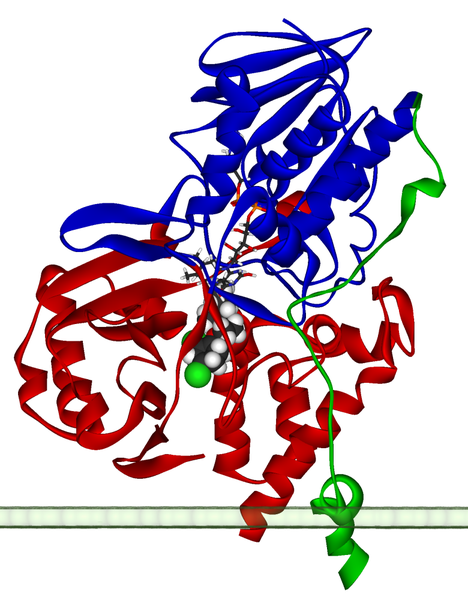
Ribbon diagram of a monomer of human monoamine oxidase A, with FAD and clorgiline bound, oriented as if attached to the outer membrane of a mitochondrion. Created using Accelrys DS Visualizer Pro 1.6 and GIMP. |
||
| Date | |||
| Source |
From the University of Michigan's Orientations of Proteins in Membranes database entry 2BXS. More information: De Colibus L, Li M, Binda C, Lustig A, Edmondson DE, Mattevi A (2005). "Three-dimensional structure of human monoamine oxidase A (MAO A): relation to the structures of rat MAO A and human MAO B". Proc. Natl. Acad. Sci. U.S.A. 102 (36): 12684–9. PMID 16129825. doi:10.1073/pnas.0505975102. |
||
| Author | Fvasconcellos 17:43, 12 May 2007 (UTC) | ||
| Permission (Reusing this file) |
|
File history
Click on a date/time to view the file as it appeared at that time.
| Date/Time | Thumbnail | Dimensions | User | Comment | |
|---|---|---|---|---|---|
| current | 17:43, 12 May 2007 | | 780 × 1,000 (310 KB) | Fvasconcellos (talk | contribs) | ==Summary== {{Information |Description=Ribbon diagram of a {{w|monomer}} of human {{w|monoamine oxidase A}}, with {{w|FAD}} and {{w|clorgiline}} bound, oriented as if attached to the {{w|outer membrane}} of a {{w|mitochondrion}}.<br>Created using [http:// |
You cannot overwrite this file.
File usage
The following 3 pages use this file:
Global file usage
The following other wikis use this file:
- Usage on ar.wikipedia.org
- Usage on ca.wikipedia.org
- Usage on cy.wikipedia.org
- Usage on en.wikipedia.org
- Usage on es.wikipedia.org
- Usage on fa.wikipedia.org
- Usage on fi.wikipedia.org
- Usage on gl.wikipedia.org
- Usage on hr.wikipedia.org
- Usage on hu.wikipedia.org
- Usage on ja.wikipedia.org
- Usage on ka.wikipedia.org
- Usage on kk.wikipedia.org
- Usage on ko.wikipedia.org
- Usage on ms.wikipedia.org
- Usage on nl.wikipedia.org
- Usage on pl.wikipedia.org
- Usage on pt.wikipedia.org
- Usage on ro.wikipedia.org
- Usage on ru.wikipedia.org
- Usage on sl.wikipedia.org
- Usage on uk.wikipedia.org
- Usage on www.wikidata.org
- Usage on zh.wikipedia.org