File:Ophiocomella ophiactoides (10.1590-1676-0611-BN-2017-0363) Figure 6.jpg
Jump to navigation
Jump to search

Size of this preview: 477 × 599 pixels. Other resolutions: 191 × 240 pixels | 382 × 480 pixels | 612 × 768 pixels | 816 × 1,024 pixels | 1,653 × 2,075 pixels.
Original file (1,653 × 2,075 pixels, file size: 693 KB, MIME type: image/jpeg)
Captions
Captions
Add a one-line explanation of what this file represents
Summary[edit]
| Description |
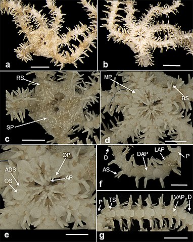
English: Figure 6. Ophiocomella ophiactoides. a, dorsal view of the animal; b, ventral view of the animal; c, dorsal view of the disk; d, ventral view of the disk; e, detail of the jaws; f, dorsal view of the arm, oriented from the distal to proximal region; g, ventral view of the arm, oriented from the proximal to the distal region. ADS, adoral shield; AP, apical papillae; AS, arm spine; BS, bursal slit; D, distal region; DAP, dorsal arm plate; LAP, lateral arm plate; MP, madreporite; OP, oral papillae; OS, oral shield; P, proximal region; RS, radial shield; SP, spines of the disk; TS, tentacle scale; VAP, ventral arm plate. Scales: a-b, d-f, 1 mm; c, g, 2 mm. |
| Date | |
| Source | https://doi.org/10.1590/1676-0611-BN-2017-0363 |
| Author | Prata, Jessica et al. Echinodermata associated to rhodoliths from Seixas Beach, State of Paraíba, Northeast Brazil. Biota Neotropica [online]. 2017, v. 17, n. 3 [Accessed 18 June 2021] , e20170363 |
| Permission (Reusing this file) |
This file is licensed under the Creative Commons Attribution 4.0 International license.
|
| Other versions |
_Figure_6_(cropped).jpg)
_Figure_6.jpg&action=edit§ion=1)
File history
Click on a date/time to view the file as it appeared at that time.
| Date/Time | Thumbnail | Dimensions | User | Comment | |
|---|---|---|---|---|---|
| current | 20:07, 18 June 2021 | | 1,653 × 2,075 (693 KB) | Christian Ferrer (talk | contribs) | {{Information | description = {{en|1=Figure 6. ''Ophiocomella ophiactoides''. a, dorsal view of the animal; b, ventral view of the animal; c, dorsal view of the disk; d, ventral view of the disk; e, detail of the jaws; f, dorsal view of the arm, oriented from the distal to proximal region; g, ventral view of the arm, oriented from the proximal to the distal region. ADS, adoral shield; AP, apical papillae; AS, arm spine; BS, bursal slit; D, distal region; DAP, dorsal arm plate; LAP, lateral a... |
You cannot overwrite this file.
File usage on Commons
The following 3 pages use this file:
_Figure_6.jpg&oldid=569895378)