File:Pharmacological-Suppression-of-CNS-Scarring-by-Deferoxamine-Reduces-Lesion-Volume-and-Increases-pone.0134371.s004.ogv
Jump to navigation
Jump to search
Size of this JPG preview of this OGG file: 800 × 600 pixels. Other resolutions: 320 × 240 pixels | 640 × 480 pixels | 1,024 × 768 pixels | 1,280 × 960 pixels | 2,560 × 1,920 pixels.
Original file (Ogg Theora video file, length 16 s, 2,560 × 1,920 pixels, 20 Mbps, file size: 38.94 MB)
Captions
Captions
Add a one-line explanation of what this file represents
Summary
[edit]| Description |
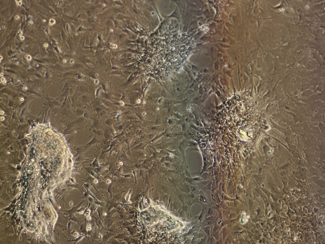
English: Live imaging of the co-cultures early after TGF. Pictures were taken from onward 10 h after TGF-β stimulation every 10th min for 8 hours. Left side: fibroblasts. Right side: astrocytes. The fibroblast layer reorganizes to form clusters that contract over time. |
||
| Date | |||
| Source | S1 Movie from Vogelaar C, König B, Krafft S, Estrada V, Brazda N, Ziegler B, Faissner A, Müller H (2015). "Pharmacological Suppression of CNS Scarring by Deferoxamine Reduces Lesion Volume and Increases Regeneration in an In Vitro Model for Astroglial-Fibrotic Scarring and in Rat Spinal Cord Injury In Vivo". PLOS ONE. DOI:10.1371/journal.pone.0134371. PMID 26222542. PMC: 4519270. | ||
| Author | Vogelaar C, König B, Krafft S, Estrada V, Brazda N, Ziegler B, Faissner A, Müller H | ||
| Permission (Reusing this file) |
This file is licensed under the Creative Commons Attribution 4.0 International license.
|
||
| Provenance |
|
File history
Click on a date/time to view the file as it appeared at that time.
| Date/Time | Thumbnail | Dimensions | User | Comment | |
|---|---|---|---|---|---|
| current | 19:30, 6 August 2015 | 16 s, 2,560 × 1,920 (38.94 MB) | Open Access Media Importer Bot (talk | contribs) | Automatically uploaded media file from Open Access source. Please report problems or suggestions here. |
You cannot overwrite this file.
File usage on Commons
The following 2 pages use this file: