File:Pseudomonas aeruginosa SEM.jpg
Jump to navigation
Jump to search

Size of this preview: 800 × 562 pixels. Other resolutions: 320 × 225 pixels | 640 × 449 pixels | 1,024 × 719 pixels | 1,280 × 899 pixels | 2,676 × 1,879 pixels.
Original file (2,676 × 1,879 pixels, file size: 785 KB, MIME type: image/jpeg)
Captions
Captions
Add a one-line explanation of what this file represents
Summary[edit]
| Description |
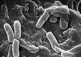
English: Scanning Electron Micrograph of Pseudomonas aeruginosa
Français : Pseudomonas aeruginosa colonise les poumons des personnes atteintes de mucoviscidose sous la forme de biofilm, qui diminue la réponse immunitaire des patients et confère à la bactérie une grande résistance aux antibiotiques. Pseudomonas aeruginosa vue au microscope électronique à balayage, est une bactérie pathogène et fréquemment rencontrée dans les infections nosocomiales.
Polski: Pałeczka ropy błękitnej w elektronowym mikroskopie skaningowym.
日本語: 緑膿菌(ムコイド型)の走査型電子顕微鏡写真菌体の表面に分泌されたムコイドが付着し、さらに一部の菌(右部)はそれに埋もれて菌全体の形が判別しにくくなっている。.
Español: Micrografía electrónica de barrido de la bacteria Pseudomonas aeruginosa, asociada con frecuencia a las infecciones pulmonares graves que complican la FQ.
Català: Micrografia electrònica d'escombrat de bacteri Pseudomonas aeruginosa, associada amb freqüència a les infeccions pulmonars greus que compliquen la FQ. |
||
| Source |
|
||
| Author |
|
||
| Permission (Reusing this file) |
PD-USGov-HHS-CDC English: None - This image is in the public domain and thus free of any copyright restrictions. As a matter of courtesy we request that the content provider be credited and notified in any public or private usage of this image. |
||
| Other versions |
|

Licensing[edit]
This image is a work of the Centers for Disease Control and Prevention, part of the United States Department of Health and Human Services, taken or made as part of an employee's official duties. As a work of the U.S. federal government, the image is in the public domain.
|
File history
Click on a date/time to view the file as it appeared at that time.
| Date/Time | Thumbnail | Dimensions | User | Comment | |
|---|---|---|---|---|---|
| current | 10:21, 4 April 2006 | | 2,676 × 1,879 (785 KB) | Y tambe (talk | contribs) | Scanning electron micrograph of '''''Pseudomonas aeruginosa''''' bacteria. Obtained from the CDC [http://phil.cdc.gov/phil/home.asp Public Health Image Library]. Image credit: CDC/Janic |
You cannot overwrite this file.
File usage on Commons
The following page uses this file:
File usage on other wikis
The following other wikis use this file:
- Usage on af.wikipedia.org
- Usage on ar.wikipedia.org
- Usage on ast.wikipedia.org
- Usage on ca.wikipedia.org
- Usage on cs.wikipedia.org
- Usage on de.wikibooks.org
- Usage on el.wikipedia.org
- Usage on en.wikipedia.org
- Usage on en.wiktionary.org
- Usage on eo.wikipedia.org
- Usage on es.wikipedia.org
- Usage on et.wikipedia.org
- Usage on eu.wikipedia.org
- Usage on fr.wikipedia.org
- Usage on gl.wikipedia.org
- Usage on id.wikipedia.org
- Usage on ja.wikipedia.org
- Usage on ka.wikipedia.org
- Usage on nl.wikipedia.org
- Usage on pl.wikipedia.org
- Usage on simple.wikipedia.org
- Usage on tr.wikipedia.org
- Usage on zh.wikipedia.org