File:Pterosaur respiratory system.jpg
(Redirected from File:Pterosaurs.jpg)

Size of this preview: 444 × 599 pixels. Other resolutions: 178 × 240 pixels | 356 × 480 pixels | 569 × 768 pixels | 759 × 1,024 pixels | 1,517 × 2,048 pixels | 3,006 × 4,057 pixels.
Original file (3,006 × 4,057 pixels, file size: 2.52 MB, MIME type: image/jpeg)
Captions
Captions
Add a one-line explanation of what this file represents
| Description |
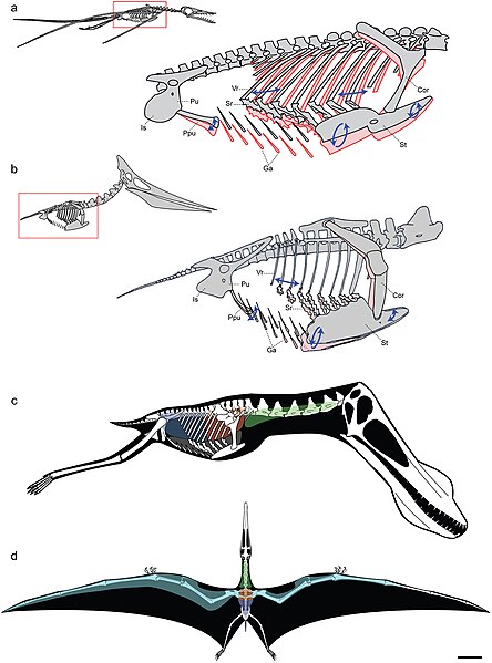
Models of ventilatory kinematics and the pulmonary air sac system of pterosaurs. a, Model of ventilatory kinematics in Rhamphorhynchus. Thoracic movement induced by the ventral intercostal musculature results in forward and outward displacement of the distal vertebral and proximal sternal ribs, and ventral displacement of the sternum, upon inspiration (blue arrows and pink outline). In addition, ventral expansion of the abdomen is induced through caudoventral rotation of the prepubis. Ranges of skeletal movement were modelled after those observed in vivo in the avian thorax and the crocodylian pelvis [26], [27]. Rhamphorhynchus modified from Wellnhofer [48]. b, Model of ventilatory kinematics in Pteranodon wherein the fused anterior vertebral ribs and articulation of the scapulocoracoid with the supraneural plate and anterior sternum limit movement of the anterior sternum, which cannot undergo elliptical rotation. However, the posterior vertebral ribs, sternal ribs, sternum, and prepubis are still capable of anterodorsal-posteroventral excursions facilitating volumetric increases and decreases of the thorax during inspiration-expiration. Pteranodon modified from Bennett [29]. c, d, reconstruction of pulmonary air sac system in the Lower Cretaceous ornithocheirid Anhanguera santanae (AMNH 22555). c, Lateral view showing the inferred position of the lungs (orange), cervical (green) and abdominal air sacs (blue), as predicted on the basis of postcranial skeletal pneumaticity. Thoracic air sacs (shown in grey) are also likely to have been present, but generally do not leave a distinct osteological trace. Humerus and more distal forelimb not shown. d, Dorsal view illustrating the inferred position of subcutaneous diverticular networks (light blue) distally along the wing. The right side depicts a conservative estimate for the size of the airsac network, limiting it to the pre-axial margin of the wing based solely on the presence of pneumatic foramina in closely positioned wing bones. The left side depicts the likely maximal size of an inferred diverticular network, accounting for its inclusion between the dorsal and ventral layers of the wing membrane. Scale = 10 cm. Skeletal reconstruction in c, d modified from Wellnhofer [49]. Abbreviations: as in figure 2, and: Cor: coracoid portion of scapulocoracoid, Ga: gastralia. |
| Date | |
| Source | http://www.plosone.org/article/info%3Adoi%2F10.1371%2Fjournal.pone.0004497;jsessionid=A57F0FDB595AC49992E2B5A390FA104C |
| Author | Leon P. A. M. Claessens, Patrick M. O'Connor, David M. Unwin |
|
This file is licensed under the Creative Commons Attribution 2.5 Generic license.
|
This file was published in a Public Library of Science journal. Their website states that the content of all PLOS journals is published under the Creative Commons Attribution 4.0 license (or its previous version depending on the publication date), unless indicated otherwise.
|
File history
Click on a date/time to view the file as it appeared at that time.
| Date/Time | Thumbnail | Dimensions | User | Comment | |
|---|---|---|---|---|---|
| current | 21:17, 2 March 2009 | | 3,006 × 4,057 (2.52 MB) | FunkMonk (talk | contribs) | {{Information |Description=Models of ventilatory kinematics and the pulmonary air sac system of pterosaurs. a, Model of ventilatory kinematics in Rhamphorhynchus. Thoracic movement induced by the ventral intercostal musculature results in forward and out |
You cannot overwrite this file.
File usage on Commons
The following 2 pages use this file:
.png)
File usage on other wikis
The following other wikis use this file:
- Usage on af.wikipedia.org
- Usage on ar.wikipedia.org
- Usage on en.wikipedia.org
- Usage on es.wikipedia.org
- Usage on it.wikipedia.org
- Usage on ko.wikipedia.org
- Usage on nl.wikipedia.org
- Usage on oc.wikipedia.org
- Usage on outreach.wikimedia.org
- Usage on pt.wikipedia.org
- Usage on ru.wikipedia.org
- Usage on tr.wikipedia.org
- Usage on vi.wikipedia.org
- Usage on zh.wikipedia.org