File:SEM micrographs (10.3897-zookeys.802.30236) Figure 4.jpg
Jump to navigation
Jump to search
Size of this preview: 427 × 599 pixels. Other resolutions: 171 × 240 pixels | 342 × 480 pixels | 547 × 768 pixels | 729 × 1,024 pixels | 1,512 × 2,122 pixels.
Original file (1,512 × 2,122 pixels, file size: 1.86 MB, MIME type: image/jpeg)
Captions
Captions
Add a one-line explanation of what this file represents
Summary[edit]
_Figure_4.jpg&action=edit§ion=1)
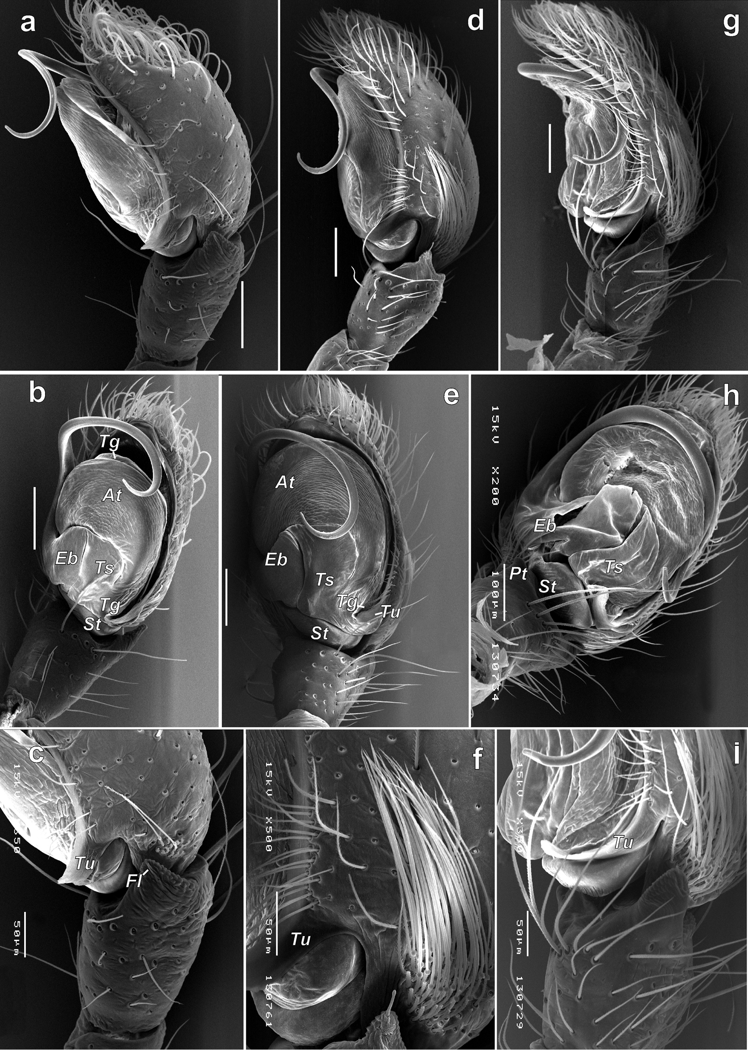
| Description | Figure 4; SEM micrographs of the male palp of Porrhoclubiona bosmansi sp. n. (a–c), P. moradmandi sp. n. (d–f) and P. leucaspis (g–i). a, d, g retrolateral b, e retro-ventral h ventral c, i tibia and proximal part of bulb and cymbium; retrolateral f cymbium and part of tegulum, retrolateral. Abbreviations: At–anterior part of tegulum, Eb–base of embolus, Fl–filamentous extension, Pt–prolateral tibial apophysis, St–subtegulum, Tg–tegular groove, Ts–sclerotised part of tegulum, Tu–tutaculum. |
| Date | |
| Source | https://doi.org/10.3897/zookeys.802.30236.figure4 (license) |
| Author | Marusik YM, Omelko MM (2018) A survey of the Porrhoclubiona Lohmander, 1944 from Central Asia (Araneae, Clubiondae). ZooKeys 802: 19-38. https://doi.org/10.3897/zookeys.802.30236 |
| Permission (Reusing this file) |
This file is licensed under the Creative Commons Attribution 4.0 International license.
|
File history
Click on a date/time to view the file as it appeared at that time.
| Date/Time | Thumbnail | Dimensions | User | Comment | |
|---|---|---|---|---|---|
| current | 14:39, 24 November 2019 | | 1,512 × 2,122 (1.86 MB) | Christian Ferrer (talk | contribs) | GWToolset: Creating mediafile for Christian Ferrer. |
You cannot overwrite this file.
File usage on Commons
There are no pages that use this file.
_Figure_4.jpg&oldid=708962473)