File:Sphaerodoropsis plurituberculata (10.11646-zootaxa.4019.1.9) Figure 5.jpg
Jump to navigation
Jump to search
Size of this preview: 405 × 599 pixels. Other resolutions: 162 × 240 pixels | 324 × 480 pixels | 519 × 768 pixels | 692 × 1,024 pixels | 1,648 × 2,439 pixels.
Original file (1,648 × 2,439 pixels, file size: 2.12 MB, MIME type: image/jpeg)
Captions
Captions
Add a one-line explanation of what this file represents
Summary
[edit]_Figure_5.jpg&action=edit§ion=1)
| Description |
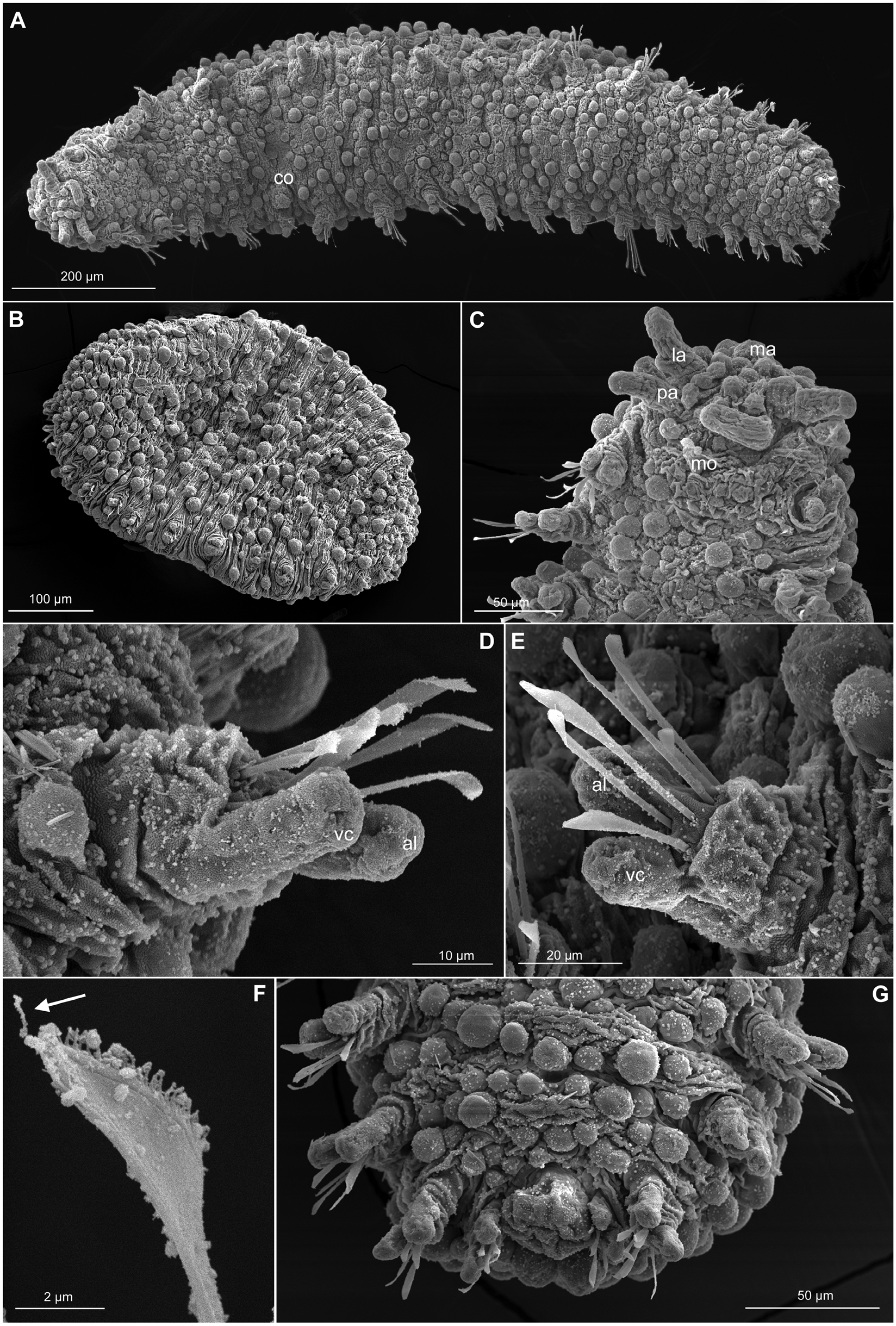
English: FIGURE 5. Sphaerodoropsis plurituberculata n. sp. SEM images. A. Male, ventral view, anterior end on left side, with copulatory organs on chaetiger 6 (co); B. Whole specimen, lateral view, anterior end on left side; C. Anterior end, ventral view; digitiform palps (pa) and lateral antennae (la), hemi-spherical median antenna (ma); mouth (mo) anterior to first chaetiger; D. Anterior chaetiger, ventral view with six chaetae, and digitiform ventral cirrus (vc) and acicular lobe (al); E. Mid-body chaetiger, posterior view; F. Detail of chaetae with conspicuous serration and distal spine (arrow); G. Posterior end with terminal pygidium. |
| Date | |
| Source | https://dx.doi.org/10.11646/zootaxa.4019.1.9 |
| Author | Capa, M. & Rouse, G.W. 2015. Sphaerodoridae (Annelida) from Lizard Island, Great Barrier Reef, Australia, including the description of two new species and reproductive notes. Zootaxa 4019 (1): 168–183. |
| Permission (Reusing this file) |
This file is licensed under the Creative Commons Attribution 3.0 Unported license.
|
File history
Click on a date/time to view the file as it appeared at that time.
| Date/Time | Thumbnail | Dimensions | User | Comment | |
|---|---|---|---|---|---|
| current | 08:11, 26 June 2021 | | 1,648 × 2,439 (2.12 MB) | Christian Ferrer (talk | contribs) | {{Information | description = {{en|1=FIGURE 5. ''Sphaerodoropsis plurituberculata'' n. sp. SEM images. A. Male, ventral view, anterior end on left side, with copulatory organs on chaetiger 6 (co); B. Whole specimen, lateral view, anterior end on left side; C. Anterior end, ventral view; digitiform palps (pa) and lateral antennae (la), hemi-spherical median antenna (ma); mouth (mo) anterior to first chaetiger; D. Anterior chaetiger, ventral view with six chaetae, and digitiform ventral cirrus... |
You cannot overwrite this file.
File usage on Commons
There are no pages that use this file.
_Figure_5.jpg&oldid=571539165)