File:TNFα-drives-mitochondrial-stress-in-POMC-neurons-in-obesity-ncomms15143-s2.ogv
Jump to navigation
Jump to search
Size of this JPG preview of this OGG file: 600 × 600 pixels. Other resolutions: 240 × 240 pixels | 480 × 480 pixels | 768 × 768 pixels | 1,024 × 1,024 pixels.
Original file (Ogg Theora video file, length 10 s, 1,024 × 1,024 pixels, 475 kbps, file size: 580 KB)
Captions
Captions
Add a one-line explanation of what this file represents
Summary
[edit]| Description |
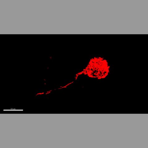
English: Supplementary Movie 1 A 3D-reconstructed neuron infected by RABVΔGMitoRFP. All mitochondria in soma and neurites were labeled by MitoRFP. Mitochondrial lengths vary from less than 0.5μm to 5μm. In the neurites, mitochondria are visible in two rails, one is supposed for anterograde transporting mitochondria from the cell body to the synaptic terminals, and the other one for the retrograde transporting mitochondria from the synaptic terminals to the cell body. |
||
| Date | |||
| Source | Video file from Yi C, Walter M, Gao Y, Pitra S, Legutko B, Kälin S, Layritz C, García-Cáceres C, Bielohuby M, Bidlingmaier M, Woods S, Ghanem A, Conzelmann K, Stern J, Jastroch M, Tschöp M (2017). "TNFα drives mitochondrial stress in POMC neurons in obesity". Nature Communications. DOI:10.1038/ncomms15143. PMID 28489068. PMC: 5436136. | ||
| Author | Yi C, Walter M, Gao Y, Pitra S, Legutko B, Kälin S, Layritz C, García-Cáceres C, Bielohuby M, Bidlingmaier M, Woods S, Ghanem A, Conzelmann K, Stern J, Jastroch M, Tschöp M | ||
| Permission (Reusing this file) |
This file is licensed under the Creative Commons Attribution 4.0 International license.
|
||
| Provenance |
|
File history
Click on a date/time to view the file as it appeared at that time.
| Date/Time | Thumbnail | Dimensions | User | Comment | |
|---|---|---|---|---|---|
| current | 21:36, 28 May 2017 | 10 s, 1,024 × 1,024 (580 KB) | Open Access Media Importer Bot (talk | contribs) | Automatically uploaded media file from Open Access source. Please report problems or suggestions here. |
You cannot overwrite this file.
File usage on Commons
The following page uses this file: