File:The-study-of-muscle-remodeling-in-Drosophila-metamorphosis-using-in-vivo-microscopy-and-bioimage-1471-2105-13-S17-S14-S1.ogv
Jump to navigation
Jump to search
Size of this JPG preview of this OGG file: 800 × 400 pixels. Other resolutions: 320 × 160 pixels | 640 × 320 pixels | 1,024 × 512 pixels.
Original file (Ogg Theora video file, length 38 s, 1,024 × 512 pixels, 1.16 Mbps, file size: 5.22 MB)
Captions
Captions
Add a one-line explanation of what this file represents
Summary
[edit]| Description |
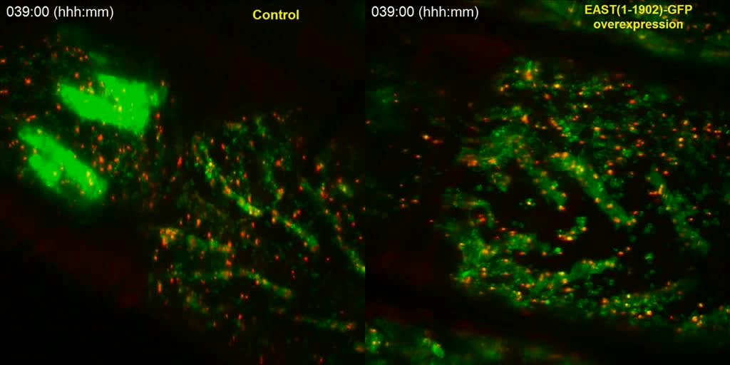
English: Temporal comparisons of time-lapse images facilitate the discovery of phenotypic abnormalities resulting from genetic perturbations. The movie shows metamorphosis of two genotypes at 30 minute intervals, a control specimen (left panel) expressing Grasp65-GFP (green) and Histone-mKO (red) and specimen expressing EAST(1-1902)-GFP (right panel), tau-GFP (green) and Histone-mKO (red). The time-lapse starts during the prepupal stage (-6 hours) and last until late pupal stage (+88 hours). The transition from the prepupal to pupal stage begins at zero hours. Note that EAST(1-1902)-GFP overexpression lead to suppression of muscle histolysis in the first 25 hours of the pupal stage and abnormalities of muscle morphology from +38 hours onwards. See Figure 3 for more details. |
||
| Date | |||
| Source | Chinta R, Tan J, Wasser M (2012). "The study of muscle remodeling in Drosophila metamorphosis using in vivo microscopy and bioimage informatics". BMC Bioinformatics. DOI:10.1186/1471-2105-13-S17-S14. PMC: 3521216. | ||
| Author | Chinta R, Tan J, Wasser M | ||
| Permission (Reusing this file) |
This file is licensed under the Creative Commons Attribution 2.0 Generic license.
|
||
| Provenance |
|
File history
Click on a date/time to view the file as it appeared at that time.
| Date/Time | Thumbnail | Dimensions | User | Comment | |
|---|---|---|---|---|---|
| current | 10:02, 28 December 2012 | 38 s, 1,024 × 512 (5.22 MB) | Open Access Media Importer Bot (talk | contribs) | Automatically uploaded media file from Open Access source. Please report problems or suggestions here. |
You cannot overwrite this file.
File usage on Commons
There are no pages that use this file.