File:Trypanosome-Motion-Represents-an-Adaptation-to-the-Crowded-Environment-of-the-Vertebrate-Bloodstream-ppat.1003023.s011.ogv
Jump to navigation
Jump to search
Size of this JPG preview of this OGG file: 800 × 500 pixels. Other resolutions: 320 × 200 pixels | 640 × 400 pixels | 1,024 × 640 pixels | 1,280 × 800 pixels | 1,728 × 1,080 pixels.
Original file (Ogg Theora video file, length 15 s, 1,728 × 1,080 pixels, 1.52 Mbps, file size: 2.73 MB)
Captions
Captions
Add a one-line explanation of what this file represents
Summary
[edit]| Description |
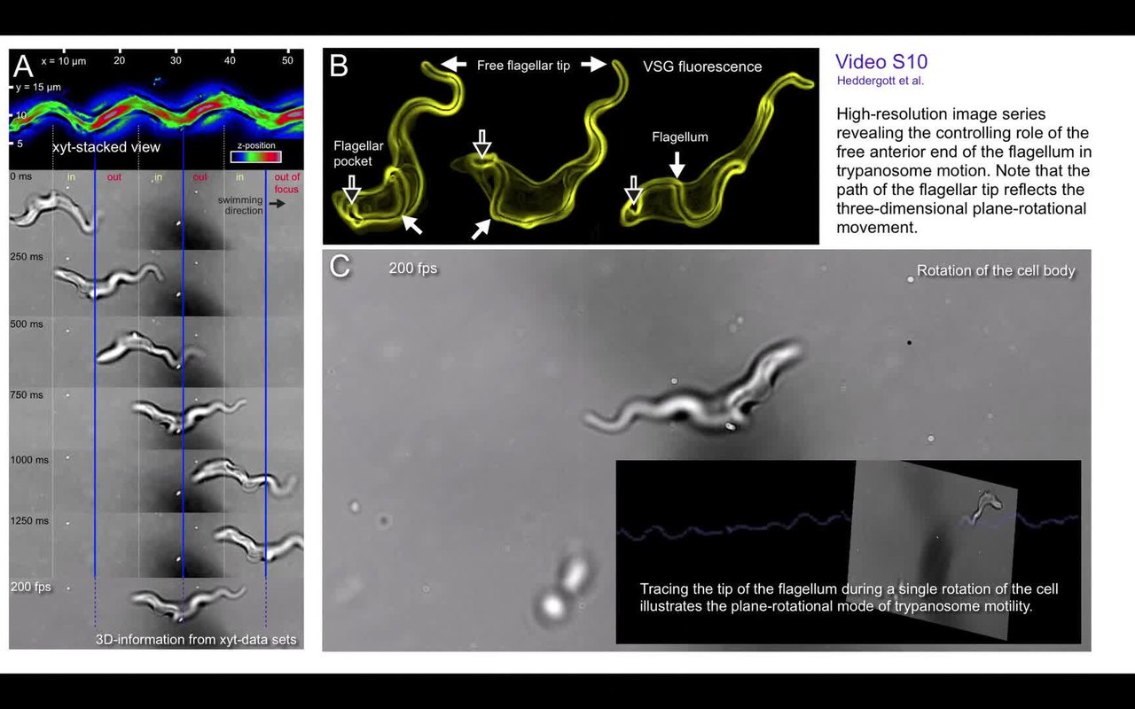
English: Out of focus information reveals three-dimensional information. (A) High speed transmitted light microscopic video corresponding to the still images in Fig. 3. The in and out of focus phases could be assigned in a periodic pattern to the projection of the cells path in order to show the helical nature of the path. In the slow motion video the rotation can be seen directly. Note the appearance and disappearance of the flagellum above or beneath the cell body, as it comes in and goes out of focus. (B) Volume-rendered models of trypanosomes, surface-labeled with AMCA-sulfo-NHS. The course of the flagellum attached to the cell body (closed arrows) is clearly visible from the flagellar pocket (open arrows) to the anterior free end. The flagellum characteristically describes a turn of about 180 degrees around the cell body, counter-clockwise in swimming direction. (C) In order to supplement the description of the three-dimensional helical path, the free anterior tip of the flagellum is traced. Note that the path of the flagellar tip along the time axis (blue) depicts a wave form that corresponds to the spatial orientation of the planar wave of the free part of the flagellum. The visible amplitude is highest when the wave lies in the viewer's xy-plane and lowest when it has rotated 90° and lies in the viewer's xz-plane. |
||
| Date | |||
| Source | Video S10 from Heddergott N, Krüger T, Babu S, Wei A, Stellamanns E, Uppaluri S, Pfohl T, Stark H, Engstler M (2012). "Trypanosome Motion Represents an Adaptation to the Crowded Environment of the Vertebrate Bloodstream". PLOS Pathogens. DOI:10.1371/journal.ppat.1003023. PMID 23166495. PMC: 3499580. | ||
| Author | Heddergott N, Krüger T, Babu S, Wei A, Stellamanns E, Uppaluri S, Pfohl T, Stark H, Engstler M | ||
| Permission (Reusing this file) |
|
||
| Provenance |
|
File history
Click on a date/time to view the file as it appeared at that time.
| Date/Time | Thumbnail | Dimensions | User | Comment | |
|---|---|---|---|---|---|
| current | 15:54, 3 December 2012 | 15 s, 1,728 × 1,080 (2.73 MB) | Open Access Media Importer Bot (talk | contribs) | Automatically uploaded media file from Open Access source. Please report problems or suggestions here. |
You cannot overwrite this file.
File usage on Commons
There are no pages that use this file.