File:Tubocurarine-from-xtal-3D-noCH-bs-17.png

Original file (3,053 × 2,343 pixels, file size: 731 KB, MIME type: image/png)
Captions
Captions
Summary[edit]
| Description |
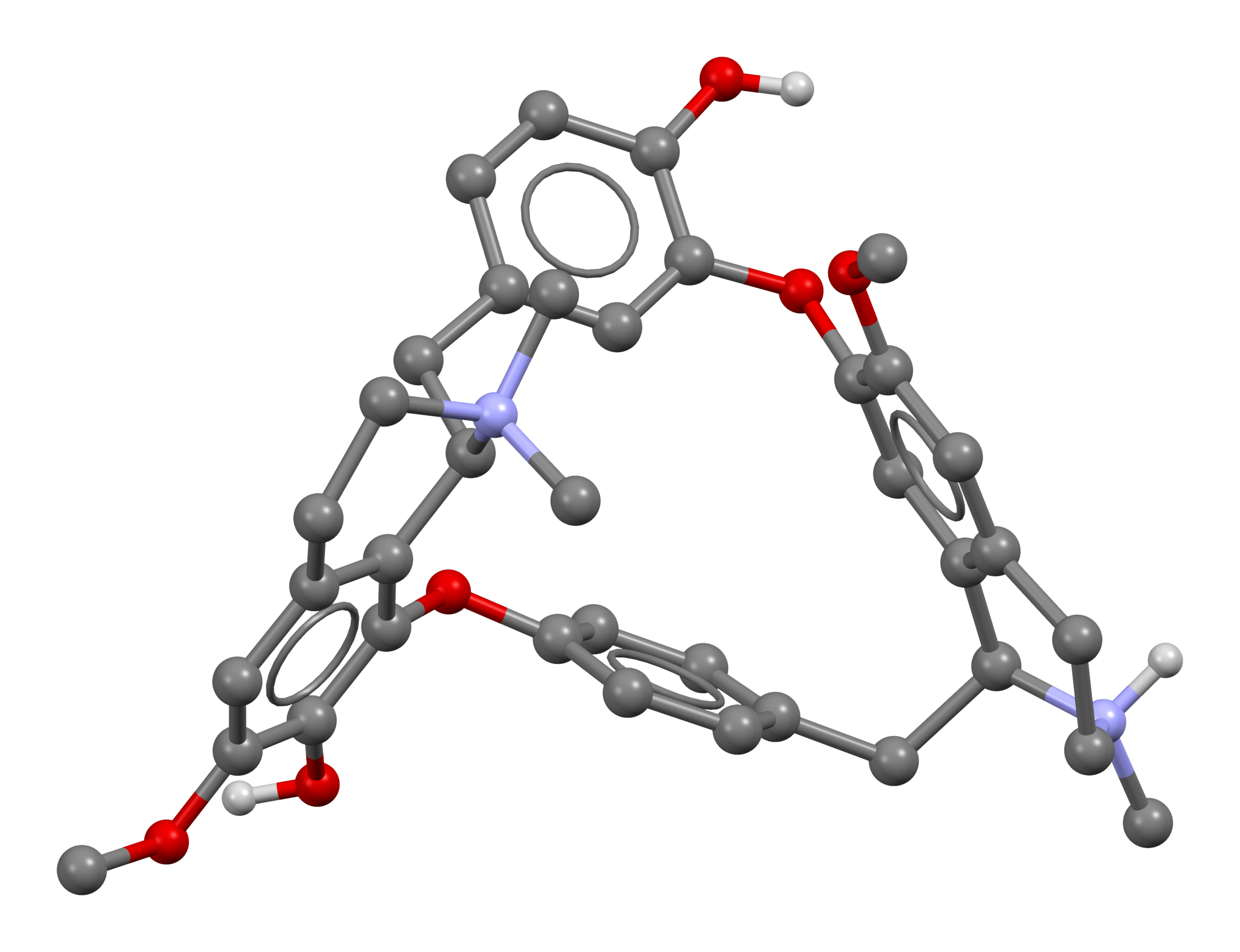
Ball-and-stick model of a tubocurarine ion, [C37H42N2O6]2+, as found in the crystal structure of D-tubocurarine dichloride pentahydrate. The structure was reported in CSD entry TUBCUR10 from Acta Cryst. B (1973) 29, 935-942. Hydrogen atoms bonded to carbon have been omitted for clarity, but hydrogens attached to heteroatoms remain. Colour code:
|
||
| Date | |||
| Source | Own work | ||
| Author | Ben Mills | ||
| Permission (Reusing this file) |
|
||
| Other versions | File:Tubocurarine-3D-sticks.png |
File history
Click on a date/time to view the file as it appeared at that time.
| Date/Time | Thumbnail | Dimensions | User | Comment | |
|---|---|---|---|---|---|
| current | 14:49, 7 April 2021 | | 3,053 × 2,343 (731 KB) | Benjah-bmm27 (talk | contribs) | == {{int:filedesc}} == {{Information | Description = Ball-and-stick model of a tubocurarine ion, [C<sub>37</sub>H<sub>42</sub>N<sub>2</sub>O<sub>6</sub>]<sup>2+</sup>, as found in the crystal structure of D-tubocurarine dichloride pentahydrate. The structure was reported in CSD entry [https://www.ccdc.cam.ac.uk/structures/Search?Ccdcid=TUBCUR10&DatabaseToSearch=Published... |
You cannot overwrite this file.
File usage on Commons
There are no pages that use this file.