File:Zebrafish Embryonic Notochord Development The pacsin3 MO phenotype.jpg

Original file (843 × 895 pixels, file size: 831 KB, MIME type: image/jpeg)
Captions
Captions
Summary
[edit]
| Description |
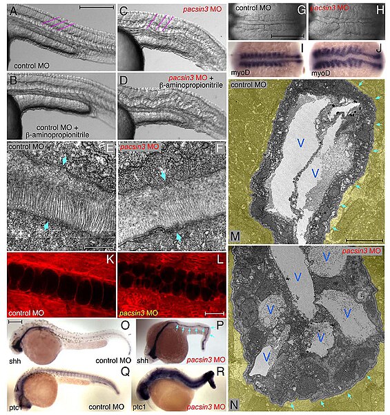
Figure 5. The pacsin3 MO phenotype. (A–D) Lateral notochord morphology in 5 ng control or pacsin3 MO-injected 24 hpf embryos treated with (B and D) or without (A and C) 10 mM b-aminoproprionitrile to disrupt the notochordal sheath. The relative angle of the normally chevron-shaped somites is indicated (purple). Anterior is left. Bar = 250 mm. (E–F) Thin section EM images of the trilaminar perinotochordal sheath in 5 ng control or pacsin3 MO-injected 24 hpf embryos. Arrows demarcate the boundary of the sheath. Bar = 0.5 mm. (G–J) Close-up dorsal views of the chordamesoderm at the 10-somite stage in 5 ng control (G and I) or pacsin3 (H and J) MO-injected embryos. Anterior is left. (I–J) Embryonic myoD mRNA localization by whole mount in situ. Bar = 250 mm. (K–L) Representative confocal sections of the lateral notochord region from BODIPY-Texas red labeled [88] live 24 hpf embryos after 5 ng control or pacsin3 MO injection. Bar = 50 mm. (M–N) Thin section EM micrographs of cross-sections through the notochord of 5 ng control or pacsin3 MO-injected 24 hpf embryos. Extra-notochord tissue is pseudocolored yellow, and vacuoles (V) and the perinotochordal sheath (arrows) are indicated. Bar = 10 mm. (O–R) Embryonic sonic hedgehog (shh) and patched1 (ptc1) mRNA localization by whole mount in situ in 5 ng control or pacsin3 MO-injected 24 hpf embryos. Bar = 250 m |
| Date | |
| Source | Edeling MA, Sanker S, Shima T, Umasankar PK, Ho¨ ning S, et al. (2009) Structural Requirements for PACSIN/Syndapin Operation during Zebrafish Embryonic Notochord Development. PLoS ONE 4(12): e8150. doi:10.1371/journal.pone.0008150 https://d-scholarship.pitt.edu/13299/1/Structural_Requirements.pdf |
| Author | James Keen, Thomas Jefferson University, United States of America |

|
This file, which was originally posted to an external website, has not yet been reviewed by an administrator or reviewer to confirm that the above license is valid. See Category:License review needed for further instructions.
|
ß 2009 Edeling et al. This is an open-access article distributed under the terms of the Creative Commons Attribution License, which permits unrestricted use, distribution, and reproduction in any medium, provided the original author and source are credited
Licensing
[edit]
- You are free:
- to share – to copy, distribute and transmit the work
- to remix – to adapt the work
- Under the following conditions:
- attribution – You must give appropriate credit, provide a link to the license, and indicate if changes were made. You may do so in any reasonable manner, but not in any way that suggests the licensor endorses you or your use.
File history
Click on a date/time to view the file as it appeared at that time.
| Date/Time | Thumbnail | Dimensions | User | Comment | |
|---|---|---|---|---|---|
| current | 10:12, 27 April 2024 | | 843 × 895 (831 KB) | Rasbak (talk | contribs) | {{Information |description=Figure 5. The pacsin3 MO phenotype. (A–D) Lateral notochord morphology in 5 ng control or pacsin3 MO-injected 24 hpf embryos treated with (B and D) or without (A and C) 10 mM b-aminoproprionitrile to disrupt the notochordal sheath. The relative angle of the normally chevron-shaped somites is indicated (purple). Anterior is left. Bar = 250 mm. (E–F) Thin section EM images of the trilaminar perinotochordal sheath in 5 ng control or pacsin3 MO-injected 24 hpf embryos.... |
You cannot overwrite this file.
File usage on Commons
There are no pages that use this file.