File:Condensation and resolution of human sister chromatids in early mitosis.svg
Jump to navigation
Jump to search

Size of this PNG preview of this SVG file: 512 × 585 pixels. Other resolutions: 210 × 240 pixels | 420 × 480 pixels | 672 × 768 pixels | 896 × 1,024 pixels | 1,792 × 2,048 pixels.
Original file (SVG file, nominally 512 × 585 pixels, file size: 934 KB)
Captions
Captions
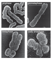
Condensation and resolution of human sister chromatids in early mitosis
Summary[edit]
| Description |
English: Scanning electron microscopy reveals that sister-chromatid pairs first condense into single rod-like structures during prophase. As mitosis proceeds, chromatid arms are gradually resolved and become almost completely distinct by the end of metaphase. Kindly provided by Adrian T. Sumner. From Sumner, A.T.: Chromosoma 1991, 100:410–418.[1] |
| Date | |
| Source | The Cell Cycle. Principles of Control. |
| Author | David O Morgan |
Licensing[edit]

|
The copyright holder of this file allows anyone to use it for any purpose, provided that the copyright holder is properly attributed. Redistribution, derivative work, commercial use, and all other use is permitted. |
|
|
File history
Click on a date/time to view the file as it appeared at that time.
| Date/Time | Thumbnail | Dimensions | User | Comment | |
|---|---|---|---|---|---|
| current | 22:28, 6 May 2020 | | 512 × 585 (934 KB) | Rob Hurt (talk | contribs) | Fixed white outlines |
| 22:18, 6 May 2020 |  | 512 × 585 (934 KB) | Rob Hurt (talk | contribs) | Uploaded a work by David O Morgan from The Cell Cycle. Principles of Control. with UploadWizard |
You cannot overwrite this file.
File usage on Commons
There are no pages that use this file.
File usage on other wikis
The following other wikis use this file:
- Usage on az.wikipedia.org
- Usage on bn.wikipedia.org
- Usage on bs.wikipedia.org
- Usage on de.wikibooks.org
- Usage on el.wikipedia.org
- Usage on en.wikipedia.org
- Usage on es.wikipedia.org
- Usage on ms.wikipedia.org
- Usage on sv.wikipedia.org
- Usage on vi.wikipedia.org