File:Metaphase meiosis II.png
Jump to navigation
Jump to search

Size of this preview: 800 × 589 pixels. Other resolutions: 320 × 235 pixels | 640 × 471 pixels | 1,024 × 753 pixels | 1,280 × 942 pixels | 1,392 × 1,024 pixels.
Original file (1,392 × 1,024 pixels, file size: 1.15 MB, MIME type: image/png)
Captions
Captions
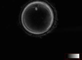
Metaphase II human oocyte.
Summary
[edit]
| Description |
Українська: Зрілий та готовий до запліднення ооцит людини. В позиції на 12 годину в перивітеліновому просторі знаходиться перше полярне тільце. Під полярним тільцем в самому ооциті чітко візуалізується веретено другого поділу мейозу на стадії метафази. Візуалізується тришарова блискуча оболонка ооциту. Поляризаційна мікроскопія. Зображення отримано на мікроскопі Nikon eclipse Ti2 (200x збільшення) з використанням системи візуалізації Oosight Meta imaging system Hamilton Thorne
English: Metaphase II human oocyte. A mature and ready for fertilization human oocyte. At the 12 o'clock position in the perivitelline space, the first polar body is located. Under the polar body in the oocyte itself, the spindle of the second division of meiosis at the metaphase stage is clearly visualized. The three layers of the zona pellucida are visible. Polarized light microscopy. The image was obtained on a Nikon eclipse Ti2 microscope (200x magnification) using the Oosight Meta imaging system Hamilton Thorne. |
| Date | |
| Source | Own work |
| Author | OlgaMaliuta |
Licensing
[edit]
I, the copyright holder of this work, hereby publish it under the following license:
This file is licensed under the Creative Commons Attribution-Share Alike 4.0 International license.
- You are free:
- to share – to copy, distribute and transmit the work
- to remix – to adapt the work
- Under the following conditions:
- attribution – You must give appropriate credit, provide a link to the license, and indicate if changes were made. You may do so in any reasonable manner, but not in any way that suggests the licensor endorses you or your use.
- share alike – If you remix, transform, or build upon the material, you must distribute your contributions under the same or compatible license as the original.
| This image was uploaded as part of Science Photo Competition 2023 in Ukraine. |

|
This media file is uncategorized.
Please help improve this media file by adding it to one or more categories, so it may be associated with related media files (how?), and so that it can be more easily found.
Please notify the uploader with {{subst:Please link images|File:Metaphase meiosis II.png}} ~~~~ |
File history
Click on a date/time to view the file as it appeared at that time.
| Date/Time | Thumbnail | Dimensions | User | Comment | |
|---|---|---|---|---|---|
| current | 23:37, 19 December 2023 | | 1,392 × 1,024 (1.15 MB) | OlgaMaliuta (talk | contribs) | Uploaded own work with UploadWizard |
You cannot overwrite this file.
File usage on Commons
The following 2 pages use this file:
File usage on other wikis
The following other wikis use this file:
- Usage on ua.wikimedia.org
- Usage on uk.wikipedia.org