Template:Histopathology of Pilomatricoma - description
Jump to navigation
Jump to search
Histopathology of pilomatricoma, high magnification, H&E stain, showing the characteristic components:
Stroma of fibrovascular connective tissue
surrounding irregularly shaped, lobulated islands containing basaloid cells (being darkly stained, round or elongated, with indistinct cell borders and minimal cytoplasm, with nuclei being round to ovoid, deeply basophilic and generally prominent nucleoli), which abruptly or (like here) gradually transitions into ghost cells (having abundant, pale, eosinophilic cytoplasm, well defined cell borders and a central clear area, but only only faint traces of nuclear material), which in turn may transition into keratinaceous to amorphous necrosis.[1]
Reference[edit]
- ↑ Punnya V Angadi (2009-06-01). Skin: Pilomatricoma. Atlas of Genetics and Cytogenetics in Oncology and Haematology.
Gallery of the same case[edit]
-
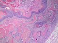 Low magnification
 -
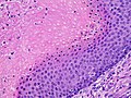 High magnification
 -
 High magnification, annotated


