Category:Antibodies
Jump to navigation
Jump to search
immune system protein  | |||||
| Upload media | |||||
| Pronunciation audio | |||||
|---|---|---|---|---|---|
| Instance of |
| ||||
| Subclass of |
| ||||
| Part of | |||||
| Manufacturer | |||||
| |||||
Subcategories
This category has the following 23 subcategories, out of 23 total.
- SVG antibodies (99 F)
?
A
C
F
H
- Helminth antibodies (2 F)
I
- Immunoglobulin therapy (7 F)
N
- Neutralizing antibodies (6 F)
P
- Protozoan antibodies (6 F)
R
S
- Secukinumab (2 F)
Media in category "Antibodies"
The following 160 files are in this category, out of 160 total.
-
 1000px-LogWachs1d.PNG 922 × 513; 32 KB
-
 136-Nanobodies antibody nanobody.tif 1,647 × 949; 4.5 MB
-
 201308 immunoglobulin.png 500 × 375; 37 KB
-
 201603 antibody.png 389 × 596; 37 KB
-
 238054 web surrogate viruses.jpg 1,440 × 1,417; 153 KB
-
 255px-Antibody-zh-cn.png 255 × 360; 14 KB
-
 2fab fc.png 432 × 432; 9 KB
-
 434px-AntibodyAR copy.png 434 × 599; 35 KB
-
 Abb1 ImmGlob.jpg 720 × 540; 31 KB
-
 Ac-toxine1.png 217 × 169; 14 KB
-
 Ac-toxine2.png 213 × 198; 23 KB
-
 Aifig2.jpg 960 × 720; 70 KB
-
 An example of a lysibody.jpg 3,119 × 915; 84 KB
-
 Anatomy of an IgG.png 1,125 × 1,235; 194 KB
-
 AngeloftheWest.jpg 1,200 × 800; 348 KB
-
 Antibody (35450606566).jpg 1,149 × 1,497; 209 KB
-
 Antibody and its corresponding antigen.svg 254 × 155; 33 KB
-
 Antibody Binding .pdf 2,000 × 1,125; 10 KB
-
 Antibody chains.jpg 332 × 500; 29 KB
-
 Antibody Effector Mechanisms.png 5,025 × 4,043; 1.93 MB
-
 Antibody IgG1 surface.png 1,845 × 1,543; 1.67 MB
-
 Antibody Isotypes.png 3,000 × 2,100; 378 KB
-
 Antibody je2-gl.png 720 × 540; 71 KB
-
 Antibody je2.png 720 × 540; 49 KB
-
 Antibody s.png 500 × 447; 6 KB
-
 Antibody Structure and Antigen Binding Regions.jpg 460 × 374; 21 KB
-
 Antibody Structure.png 602 × 292; 5 KB
-
 Antibody-tr.jpg 255 × 360; 14 KB
-
 Antibody-zh.png 251 × 348; 14 KB
-
 Antibody.JPG 338 × 312; 57 KB
-
 Antibody.png 255 × 360; 13 KB
-
 Antibody1.jpg 248 × 263; 51 KB
-
 Antibody1.JPG 389 × 263; 10 KB
-
 AntibodyAR.png 787 × 1,086; 46 KB
-
 AntibodyFcAR.png 1,294 × 1,299; 48 KB
-
 Antibodys in cancer therapy 01.png 2,345 × 1,726; 484 KB
-
 AntibodyX.JPG 492 × 362; 20 KB
-
 Anticorps en rotation.gif 144 × 144; 83 KB
-
 Anticorps schema.png 20 × 20; 239 bytes
-
 Anticorps.png 272 × 274; 25 KB
-
 Anticuerpo (Antibody) (35795301294).jpg 1,149 × 1,873; 314 KB
-
 Antigen-antibody-complex.png 395 × 307; 53 KB
-
 Antigen-Antikoerper-Reaktion.jpg 794 × 248; 28 KB
-
 Antigorputza.png 794 × 1,123; 128 KB
-
 Antikörper-Hybride.png 795 × 288; 29 KB
-
 Antilichaam.svg 400 × 340; 13 KB
-
 B-cell receptor structure ku.png 773 × 646; 96 KB
-
 Basic Immunoglobuline 2.jpg 283 × 283; 35 KB
-
 Biochem-polyclonal production.jpg 1,214 × 705; 78 KB
-
 Biochem-polyclonal.jpg 679 × 353; 32 KB
-
 Cadeia Leve e Cadeia Pesada das Imunoglobulinas..png 1,204 × 1,031; 567 KB
-
 Cambio clase recombinacion.PNG 750 × 750; 9 KB
-
 Captura de pantalla MZ4.png 332 × 197; 26 KB
-
 Capture ab.png 642 × 535; 100 KB
-
-
 Class switch recombination gl.png 750 × 750; 19 KB
-
 Class switch recombination.png 750 × 750; 9 KB
-
 Comp B.jpg 156 × 237; 31 KB
-
 Complement Pathways 2 - closer look.png 4,409 × 3,274; 1.12 MB
-
 Complement Pathways.png 3,000 × 2,626; 371 KB
-
-
De-Antikörper.ogg 1.5 s; 16 KB
-
De-Immunglobulin.ogg 2.6 s; 25 KB
-

-
 Diagnostic Medical Dipstick.png 2,133 × 1,600; 83 KB
-
 Diagnostics-10-00453-g001.webp 3,590 × 1,991; 1.58 MB
-
 Diagnostics-10-00453-g002.webp 2,995 × 2,765; 1.26 MB
-
 Diagnostics-10-00453-g003.webp 2,512 × 3,536; 1.53 MB
-
 Different Blood Types.png 815 × 394; 37 KB
-
 Digibind.jpg 2,931 × 2,833; 1.12 MB
-
-
 Dorsal Root Galglia Neurons.tif 6,144 × 1,920; 8.55 MB
-
 DVD-Ig Knob in Hole.png 657 × 390; 29 KB
-
 ELISPOT-en.png 517 × 404; 36 KB
-
 Epitope-mapping illustration-6-copy.png 1,309 × 855; 456 KB
-
 Epitope-mapping-MOA.jpg 567 × 322; 14 KB
-
 Epitopos.png 512 × 373; 27 KB
-
 Epitopos2.png 512 × 373; 49 KB
-
 Esquema classificació anticardiolipina..jpg 820 × 458; 65 KB
-
 Estructura pentamèrica formada per cinc IgM unides amb una cadena J.jpg 1,452 × 1,323; 180 KB
-
 F ab2 pFc.png 432 × 432; 14 KB
-
 F(ab')2 fcfragment.png 589 × 622; 40 KB
-
 Fab fcfragment colors.png 768 × 720; 38 KB
-
 Fab fcfragment.png 768 × 720; 45 KB
-
 FAM166C Antibody Staining.png 1,415 × 1,401; 2.75 MB
-
 Figure 42 02 06.jpg 544 × 586; 134 KB
-
Filamins-Regulate-Cell-Spreading-and-Initiation-of-Cell-Migration-pone.0007830.s002.ogv 8.4 s, 502 × 314; 537 KB
-
Filamins-Regulate-Cell-Spreading-and-Initiation-of-Cell-Migration-pone.0007830.s003.ogv 8.3 s, 502 × 314; 476 KB
-
Filamins-Regulate-Cell-Spreading-and-Initiation-of-Cell-Migration-pone.0007830.s004.ogv 8.1 s, 502 × 314; 460 KB
-
Filamins-Regulate-Cell-Spreading-and-Initiation-of-Cell-Migration-pone.0007830.s005.ogv 8.3 s, 590 × 408; 462 KB
-
Filamins-Regulate-Cell-Spreading-and-Initiation-of-Cell-Migration-pone.0007830.s006.ogv 7.9 s, 362 × 254; 128 KB
-
 Fox2-Fox3.png 1,000 × 909; 1.76 MB
-
 Functional.jpg 640 × 400; 15 KB
-
 Funktionsweise von therapeutischen Antikörpern.webp 1,347 × 999; 75 KB
-
 General Effector Mechanisms of B and T Cells.png 3,300 × 1,650; 554 KB
-
 Glass bottle with immunoglobulin to fight against chickenpox Wellcome L0057971.jpg 4,787 × 3,329; 3.04 MB
-
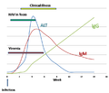 HAV Infection.png 719 × 619; 35 KB
-
 Hcp-0.0-CRYSVITA-antibody rotating 03.gif 372 × 345; 2.26 MB
-
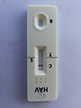 Hepatitis A Virus Spot Test.jpg 6,000 × 8,000; 9.45 MB
-
 Hipotesistrasposon.JPG 969 × 1,280; 114 KB
-
 HLA+Gliadin 1S9V.png 1,779 × 1,533; 774 KB
-
 IgA antibody.tif 1,280 × 720; 299 KB
-
 IgG1 vs IgG4 configuration.png 376 × 242; 24 KB
-
 IgM IgE scheme.png 388 × 372; 30 KB
-
 ImmunfluoreszenzI.jpg 400 × 482; 19 KB
-
 Immunoassay strip test for a cucurbit virus.gif 120 × 160; 139 KB
-
 Immunoglobineketens.png 664 × 1,000; 63 KB
-

-
 Immunoglobulin 017.jpg 1,768 × 1,326; 356 KB
-
 Immunoglobulin 018.jpg 3,072 × 2,304; 1.08 MB
-
 Immunoglobulin 019.jpg 3,072 × 2,304; 1.49 MB
-
 Immunoglobulin.png 500 × 375; 38 KB
-
 Immunohistochemistry flow chart.jpg 900 × 312; 115 KB
-
 Immunometric Assay.jpg 151 × 243; 30 KB
-
 Immuunrespons.svg 500 × 440; 1.2 MB
-
 Inmunoglobulina.png 340 × 280; 118 KB
-
 Inmunoglobulinas-estructura.png 869 × 359; 257 KB
-
 Inmunoglobulinas-tipos.png 970 × 494; 95 KB
-
 Intact antibodies and a variety of antibody fragments.png 942 × 806; 601 KB
-
 Mono-und-Polymere-zh.png 242 × 243; 20 KB
-
 Monoclonal antibodies4.jpg 1,350 × 900; 624 KB
-
 Monoclonal.png 284 × 289; 26 KB
-
 MonoclonalAb.jpg 427 × 285; 27 KB
-
 NiosomeALOK.JPG 360 × 233; 14 KB
-
 Nomenclature.png 890 × 362; 72 KB
-
 Orientación de anticuerpos en un soporte.png 1,908 × 670; 506 KB
-

-
 PDB 5ZIA cartoon and coarse surface chain instance DL20221121.tif 4,370 × 2,067; 3.88 MB
-
 Peroxisome .jpg 1,280 × 960; 381 KB
-
 Peroxisome in rat neonatal cardiomyocyte.jpg 1,280 × 960; 79 KB
-
 Polyclonal.png 274 × 263; 31 KB
-
 Ppat.1008735.g002.png 2,244 × 1,496; 654 KB
-
 Primary secondary antibody.png 712 × 632; 36 KB
-
 Recombinación VDJ.PNG 750 × 750; 8 KB
-
 Recombination-IgM-IgG1-IgG4.png 816 × 287; 33 KB
-
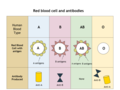 Red Blood Cell and Antibodies.png 960 × 720; 81 KB
-
 Regiões Variáveis e Constantes.png 1,003 × 942; 315 KB
-
 Schematic illustration of the TMEM81 peptide.png 1,582 × 280; 61 KB
-
 Single domain antibody.tif 1,500 × 937; 4.05 MB
-
 SMH wiki v2.png 1,904 × 2,132; 217 KB
-
 SMH wiki.png 1,836 × 2,376; 86 KB
-
 Somatic hypermutation v5.png 3,808 × 4,264; 89 KB
-
 Structure of the intact human IgG B12 antibody (PDB 1hzh).png 1,128 × 1,302; 855 KB
-
 T independent B cell Activation.png 4,844 × 2,018; 674 KB
-
 The twin brothers.tif 10,836 × 10,011; 21.31 MB
-
-
 Transition State Analog Selection Approach.png 2,665 × 1,467; 135 KB
-
 V(D)J-recombinatie.svg 800 × 700; 310 KB
-
 VDJ recombination gl.png 750 × 750; 15 KB
-
 VDJ recombination.png 750 × 750; 7 KB
-
 Η δομή της ανοσοσφαιρίνης IgG.JPG 410 × 375; 17 KB
-
 Молекулярный механизм V(D)J-реаранжировки.png 1,654 × 1,250; 399 KB
-
 Организация генетических локусов иммуноглобулина в геноме человека..png 1,181 × 570; 101 KB
-

-
 Реаранжировка и сплайсинг мРНК гена IgH.png 709 × 341; 56 KB
-
 Сигнальные последовательности – recombination signal sequence (RSS).png 1,654 × 948; 152 KB
-
 Структура иммуноглобулина и Т-клеточного рецептора.png 1,181 × 669; 127 KB
-
 ప్రతిరక్షకము ( Immunoglobulin ).png 2,048 × 2,048; 248 KB
-
 ആന്റിബോഡി തൻമാത്രയുടെ ഭാഗങ്ങൾ.png 1,749 × 1,241; 92 KB
-
 ഇമ്മ്യൂണോഗ്ലോബുലിൻ ത്രിമാനഘടന.jpg 1,235 × 631; 115 KB


.jpg)


_(35795301294).jpg)


;_5_-_granule;_6_-_mastocit;_7_-_novoformirani_medijatori_(prostaglandini,_leukotrieni).png)


2_fcfragment.png)


_antibodies_binding_to_adjacent_antigen_epitopes_on_the_surface_of_bacterial_cells..png)


.png)


J-recombinatie.svg)


J-%D1%80%D0%B5%D0%B0%D1%80%D0%B0%D0%BD%D0%B6%D0%B8%D1%80%D0%BE%D0%B2%D0%BA%D0%B8.png)


.png)

.png)

