Category:Caenorhabditis elegans
Jump to navigation
Jump to search
Domain: Eukaryota • Regnum: Animalia • Subregnum: Eumetazoa • Cladus: Bilateria • Superphylum: Protostomia • (Unranked): Ecdysozoa • Phylum: Nematoda • Classis: Chromadorea • Ordo: Rhabditida • Familia: Rhabditidae • Genus: Caenorhabditis • Species: Caenorhabditis elegans
Wikispecies has an entry on:
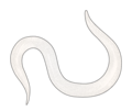
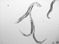
free-living species of nematode  micrografia d'un adult de C. elegans | |||||||||||||||||||||||||
| Upload media | |||||||||||||||||||||||||
| Instance of | |||||||||||||||||||||||||
|---|---|---|---|---|---|---|---|---|---|---|---|---|---|---|---|---|---|---|---|---|---|---|---|---|---|
| Significant event |
| ||||||||||||||||||||||||
| |||||||||||||||||||||||||
| |||||||||||||||||||||||||
| Taxon author | Émile Maupas, 1900 | ||||||||||||||||||||||||
| |||||||||||||||||||||||||
Subcategories
This category has the following 6 subcategories, out of 6 total.
?
C
E
S
V
Pages in category "Caenorhabditis elegans"
This category contains only the following page.
Media in category "Caenorhabditis elegans"
The following 114 files are in this category, out of 114 total.
-
 ''Caenorhabditis elegans'' intestine infected with ''Bacillus thuringiensis''.jpg 1,754 × 1,275; 895 KB
-
 2007 May.tif 615 × 615; 584 KB
-
 201108 nematode.png 465 × 392; 80 KB
-
 202205 Nematode.svg 512 × 640; 392 KB
-
 202210 nematode 2-fold stage.svg 1,000 × 1,000; 348 KB
-
 202210 nematode 3-fold stage.svg 1,000 × 1,000; 358 KB
-
 202210 nematode adult female.svg 1,000 × 1,000; 381 KB
-
 202210 nematode adult male.svg 1,000 × 1,000; 367 KB
-
 202210 nematode copulation.svg 1,000 × 1,000; 397 KB
-
 202210 nematode dauer larva formation.svg 1,000 × 1,000; 422 KB
-
 202210 nematode dumpy mutant.svg 1,000 × 1,000; 452 KB
-
 202210 nematode egg laying-defective.svg 1,000 × 1,000; 445 KB
-
 202210 nematode embryo 10 cells stage.svg 1,000 × 1,000; 294 KB
-
 202210 nematode embryo 2 cells stage.svg 1,000 × 1,000; 277 KB
-
 202210 nematode embryo 28 cells stage.svg 1,000 × 1,000; 336 KB
-
 202210 nematode embryo comma stage.svg 1,000 × 1,000; 342 KB
-
 202210 nematode long mutant.svg 1,000 × 1,000; 462 KB
-
 202210 nematode multiple vulval protrusions.svg 1,000 × 1,000; 480 KB
-
 202210 nematode on Nematoed Growth Medium petri plate.svg 1,000 × 1,000; 583 KB
-
 202210 nematode roller blister mutant.svg 1,000 × 1,000; 501 KB
-
 202210 nematode roller mutant.svg 1,000 × 1,000; 488 KB
-
 202210 nematode small mutant.svg 1,000 × 1,000; 470 KB
-
 Adult Caenorhabditis elegans.jpg 6,424 × 2,113; 737 KB
-
 ALS animal models, photos only.jpg 714 × 177; 93 KB
-
 ALS animal models.jpg 1,219 × 1,280; 122 KB
-
 ALS Disease Pathology and Proposed Disease Mechanisms.jpg 1,052 × 1,280; 194 KB
-
 Anatomía de un nematodo macho-es.svg 1,444 × 2,210; 668 KB
-
 Apical Constriction.jpg 242 × 300; 80 KB
-
 Apoptosis C. elegans hy.svg 1,877 × 387; 462 KB
-
 Apoptosis C. elegans.svg 1,877 × 387; 277 KB
-
 Apoptotic Phagocytosis in C. elegans.jpg 1,776 × 1,335; 454 KB
-
 Archives de zoologie expérimentale et générale (1900) (20139214309).jpg 1,862 × 2,964; 583 KB
-
 C elegans apoptose.png 976 × 36; 12 KB
-
 C elegans DIC s.jpg 640 × 800; 97 KB
-
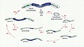
 C elegans life cycle.svg 888 × 490; 13 KB
-
 C-elegans-Schematic-drawing-of-the-two-gonads-and-uterus-of-C.jpg 600 × 314; 57 KB
-
 C. elegans body and muscle mapping.png 2,346 × 1,906; 4.05 MB
-
 C. elegans culture s.jpg 142 × 106; 25 KB
-
 C. elegans diagram.png 1,366 × 768; 136 KB
-
 C. elegans we looked in class.jpg 3,024 × 4,032; 607 KB
-
C. elegans with pencil for scale.webm 42 s, 1,024 × 768; 6.53 MB
-
 C. elegans, model organism in life sciences (28703152561).jpg 2,560 × 1,920; 369 KB
-
 C. elegns DIC l.jpg 1,392 × 1,040; 86 KB
-
 Caenorhabditis elegans (C. elegans) clip art.png 1,210 × 396; 75 KB
-
 Caenorhabditis elegans (C. elegans) clip art.svg 363 × 119; 5 KB
-
 Caenorhabditis elegans hermaphrodite adult-cs.svg 792 × 380; 2 MB
-
 Caenorhabditis elegans Oil-Red-o.tif 2,040 × 1,536; 8.97 MB
-
 CED-9 apoptosis.png 374 × 1,006; 36 KB
-
 Celegans wt nhr80rnai.png 996 × 392; 315 KB
-
 CelegansGoldsteinLabUNC 2.jpg 350 × 297; 22 KB
-
 CelegansGoldsteinLabUNC.jpg 350 × 297; 40 KB
-
 Ciclocelegans.jpg 1,280 × 720; 125 KB
-
 Complete cell lineage of C elegans.png 1,110 × 576; 41 KB
-
 CyberElegans Project.png 2,459 × 924; 2.43 MB
-
 Dosage compensation in C. elegans.png 1,487 × 1,126; 454 KB
-
 Electron micrograph of ''Caenorhabditis elegans''.jpg 213 × 213; 9 KB
-
 Examples of miRNA stem-loops.jpg 476 × 180; 41 KB
-
 Exogenous RNAi Pathway in C. elegans, edited.svg 675 × 778; 60 KB
-

-

-
 Fig 2. Gráfico del linaje celular de C. elegans..jpg 594 × 321; 28 KB
-
 Fig 3. Proceso de gastrulación en el nemátodo C.elegans..png 1,278 × 427; 121 KB
-
 Fig. 3. Mecanismo de polarización celular de C .elegans..png 720 × 540; 114 KB
-
 Fig. 3.. Mecanismo de polarización celular de C .elegans..png 720 × 540; 113 KB
-
 Fluorescent C. elangs D. melanogaster S. pombe.jpg 2,100 × 1,500; 560 KB
-
 Glycosylation.jpg 1,800 × 1,199; 305 KB
-
 IFTcelegans.JPG 1,421 × 900; 145 KB
-
 IFTcilia.jpg 1,421 × 900; 439 KB
-
 Illustration of C. elegans response against exogeneous dsRNA.png 1,048 × 601; 96 KB
-
 Main Mechanisms of Dosage Compensation.png 1,500 × 708; 173 KB
-
 Male Mating.gif 200 × 150; 3.11 MB
-
 Mecanismo de polarización celular de C .elegans..png 1,060 × 516; 61 KB
-
 Mermis nigrescens (right),Caenorhabditis elegans (left) body wall muscle.jpg 2,801 × 1,429; 551 KB
-
 Mt-tRNA(Tyr).png 2,124 × 867; 54 KB
-
 Nematode (Caenorhabditis elegans).png 465 × 380; 80 KB
-

-
 Nematode C. elegans Anatomy Relating to the Control of Meiotic Maturation.jpg 1,800 × 2,281; 2.41 MB
-

-
 Nematode C. elegans Distributions of the MES proteins in 2-cell embryos.jpg 1,652 × 816; 487 KB
-

-

-

-

-
 Nematode C. elegans germline chromatin hermaphrodite and male X chromosomes.jpg 1,400 × 1,400; 812 KB
-
 Nematode C. elegans germline chromatin.jpg 1,728 × 1,920; 766 KB
-
 Nematode C. elegans gonadogenesis.jpg 1,792 × 1,193; 557 KB
-
 Nematode C. elegans interactions in the hermaphrodite Sperm competition.jpg 1,120 × 1,368; 483 KB
-

-
 Nematode C. elegans meiosis meiotic recombination.jpg 1,800 × 1,551; 697 KB
-
 Nematode C. elegans meiosis Synaptonemal complex.jpg 1,772 × 1,422; 1.4 MB
-

-
 Nematode C. elegans Oocyte Meiotic Maturation and Egg Activation.jpg 1,744 × 1,240; 407 KB
-
 Nematode C. elegans Proteins present in P granules.png 1,179 × 836; 176 KB
-
Nematode C. elegans spermatid budding1.ogv 3.1 s, 276 × 275; 179 KB
-
 Nematode C. elegans spermatogenesis.jpg 1,800 × 1,341; 632 KB
-
 Nematode C. elegans Stages of wild type spermatogenesis.jpg 1,793 × 724; 522 KB
-
 Nematode C. elegans The early embryonic lineage in 4-cell embryos.jpg 1,312 × 1,264; 290 KB
-

-
Nematode C. elegans Time-lapse Video of Oocyte Meiotic Maturation and Ovulation.ogv 45 s, 180 × 120; 208 KB
-
 Nematode cell cycle patterns.jpg 769 × 1,604; 401 KB
-
 Nematode Diagram of meiotic events during oogenesis in the C. elegans germ line.jpg 1,800 × 1,190; 740 KB
-
 Nematode embryonic polarity.jpg 1,800 × 1,156; 679 KB
-
 Nematode P granules.jpg 886 × 1,721; 411 KB
-
 Nematode Variations in early cleavage pattern.jpg 1,800 × 1,569; 1,011 KB
-
 Nematode Variations in gastrulation.jpg 1,800 × 1,909; 1.74 MB
-
 Nematode.jpg 5,472 × 3,648; 4.37 MB
-
 Section of c elegans.jpg 1,081 × 664; 243 KB
-
 Sensory cilium.jpg 649 × 296; 40 KB
-
 Structure and signaling mechanisms of the C. elegans GSC niche..jpg 650 × 297; 77 KB
-
 Varbussid.jpg 2,316 × 1,650; 347 KB
-
 Wormandwaterbear.jpg 189 × 495; 50 KB
-
 Z curve of C.elegans chromosome III.png 640 × 480; 5 KB
-
 Инсулин-IGF сигнальный каскад в C. elegans контролирует переход в dauer форму.png 3,022 × 2,264; 216 KB
-
 Продукты daf-генов и долголетие - C. elegans.png 986 × 715; 421 KB


_(20139214309).jpg)


.jpg)


,Caenorhabditis_elegans_(left)_body_wall_muscle.jpg)
.png)


_clip_art.png)
_clip_art.svg)
.png)