Category:Lipid bilayers
Jump to navigation
Jump to search
A thin polar membrane made of two layers of lipid molecules | |||||
| Upload media | |||||
| Subclass of | |||||
|---|---|---|---|---|---|
| |||||
Subcategories
This category has the following 8 subcategories, out of 8 total.
Media in category "Lipid bilayers"
The following 75 files are in this category, out of 75 total.
-
 "Membrane Thingy"-Max 8-5-22.png 790 × 311; 90 KB
-
 A lipid micelle.svg 266 × 266; 13 KB
-
 Annular Gap Junction Vesicle.jpg 205 × 153; 61 KB
-
 Asymmetry of lipid bilayer.jpg 800 × 450; 33 KB
-
 Bilayer AFM schematic.png 2,391 × 1,503; 1.43 MB
-
 Bilayer hydration profile cn.svg 512 × 501; 130 KB
-
 Bilayer hydration profile gl.svg 648 × 634; 452 KB
-
 Bilayer hydration profile-es.svg 648 × 634; 389 KB
-
 Bilayer hydration profile.svg 648 × 634; 355 KB
-
 Bilayer scheme 2.jpg 546 × 313; 128 KB
-
 Bilayer scheme with lateral water.jpg 633 × 313; 84 KB
-
 Bilayer scheme.svg 290 × 92; 168 KB
-
 Biogènesi.jpg 1,141 × 2,129; 592 KB
-
 Brush PEG-SLB generation method.png 575 × 845; 557 KB
-
 Energized Lecitin Membrane.svg 1,052 × 744; 1.2 MB
-
 Fick's Law for gas-exchange surface.png 1,090 × 260; 33 KB
-
 Figure 03 03 09.jpg 544 × 289; 136 KB
-
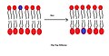 Flip Flop Diffusion.jpg 1,603 × 823; 143 KB
-
 Fluid mosaic model.jpg 640 × 400; 26 KB
-
 Fluidità membrana.jpg 960 × 720; 92 KB
-
 Hopanoid-membrane.png 3,579 × 1,952; 2.44 MB
-
 Hydrophilic surface FSL coating.svg 294 × 273; 4.12 MB
-
 Koded biomembrane.svg 1,052 × 744; 2.29 MB
-
 Lecitin-gradient-small.svg 350 × 600; 760 KB
-
 Lipid bilayer and micelle svg.png 530 × 266; 66 KB
-
 Lipid bilayer and micelle.png 530 × 266; 5 KB
-
 Lipid bilayer and micelle.svg 530 × 266; 33 KB
-
 Lipid bilayer fluid.JPG 539 × 368; 39 KB
-
 Lipid bilayer fluid.svg 459 × 387; 27 KB
-
 Lipid bilayer section.gif 300 × 195; 124 KB
-
 Lipid Bilayer.jpg 720 × 576; 87 KB
-
 Lipid Molecules (5941036454).jpg 406 × 629; 401 KB
-
 Lipid Raft.png 444 × 271; 110 KB
-
 Lipid unsaturation effect pt.svg 444 × 299; 210 KB
-
 Lipid unsaturation effect-ar.jpg 1,479 × 996; 811 KB
-
 Lipid unsaturation effect.svg 444 × 299; 159 KB
-
 Lipid-News-Kraft-5-inch-wide.jpg 1,500 × 772; 114 KB
-
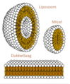 Liposoom.png 816 × 984; 437 KB
-
 Lyssaviirus.png 845 × 670; 160 KB
-
 Membran1.gif 462 × 88; 4 KB
-
 Membrane bilayer.jpg 585 × 460; 35 KB
-
 Membrane fusion via stalk formation.jpg 1,096 × 202; 25 KB
-
 Membrane orbit animated.gif 256 × 192; 2.16 MB
-
 Micella bicapa.png 400 × 250; 177 KB
-
 Micelle fosfolipidi.jpg 960 × 720; 108 KB
-
 Microdomínio de membrana (balsa lipídica).tif 448 × 286; 100 KB
-
 Mitochondrial membrane and MitoQ.jpg 924 × 921; 91 KB
-

-
 PalmitoylationAnkGc pl.jpg 340 × 360; 76 KB
-
 Peptidoglycan-membrane.png 2,083 × 1,458; 197 KB
-
 Phosphoglycerolipide.pdf 1,800 × 1,054; 117 KB
-
 Phospholipid TvanBrussel.edit.jpg 614 × 437; 167 KB
-
 Phospholipid TvanBrussel.jpg 640 × 442; 126 KB
-
 Phospholipidbyjj.jpg 1,152 × 648; 37 KB
-
 Phospholipids aqueous solution structures-ar.jpg 4,600 × 5,650; 7.52 MB
-
 Protein crowding membrane bending.png 908 × 650; 125 KB
-
 Repolarization of a Nerve Impulse.svg 512 × 382; 262 KB
-
 Schemes of macro-molecular structures peptide amphiphiles.jpg 592 × 918; 81 KB
-
 Small bilayer.png 205 × 69; 14 KB
-
 Supported bilayer.svg 635 × 288; 84 KB
-
 Supported Lipid Bilayer and Nanoparticles AFM-es.png 1,220 × 1,003; 251 KB
-
 Supported Lipid Bilayer and Nanoparticles AFM.png 1,220 × 1,003; 807 KB
-
 T-BLM.png 1,138 × 512; 17 KB
-
 The lipid and lipid bilayer-el.svg 375 × 442; 29 KB
-
 The lipid and lipid bilayer.png 300 × 354; 17 KB
-
 The membirain thingy.png 405 × 165; 39 KB
-
 Thermoreception 1.png 4,236 × 2,812; 1.12 MB
-
 Thermoreception 2.png 4,236 × 2,812; 1.18 MB
-
 Unidade de membrana.jpg 606 × 296; 80 KB
-
 Valinomycin transport.png 1,330 × 1,313; 467 KB
-
 Схематичне зображення ліпоплексу.JPG 436 × 349; 44 KB
-
 リン脂質の基本構造(ぱた).png 522 × 171; 7 KB
-
 リン脂質二重層膜の基本構造(byパタ).png 459 × 165; 13 KB
-
 二重層・リポソーム・ミセル.png 338 × 409; 133 KB
-
 細胞二重膜の模式図.svg 1,200 × 750; 137 KB


.jpg)


.png)
.png)