Category:Green fluorescent proteins
Salti al navigilo
Salti al serĉilo
protein that converts blue and ultraviolet light ranges to green light | |||||
| Alŝuti plurmedion | |||||
| Estas |
| ||||
|---|---|---|---|---|---|
| Subaro de |
| ||||
| |||||
Dosieroj en kategorio “Green fluorescent proteins”
La jenaj 200 dosieroj estas en ĉi tiu kategorio, el 223 entute.
(antaŭa paĝo) (sekva paĝo)-
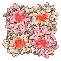 174-GFPLikeProteins 1g7k.tif 854 × 844, 2 paĝoj; 2,1 MB
-
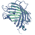
 174-GFPLikeProteins GFP-like Proteins.tif 1 656 × 1 878, 2 paĝoj; 8,93 MB
-
 202404 Green Fluorescent Protein.svg 400 × 400; 121 KB
-
-
-
-
-
-
-
-
-
-
An-actin-dependent-spindle-position-checkpoint-ensures-the-asymmetric-division-in-mouse-oocytes-ncomms8784-s4.ogv 10 s, 1 024 × 1 024; 1,05 MB
-
-
-
 Anthrax and gfp s.jpg 400 × 218; 115 KB
-
Association-of-the-Hermansky-Pudlak-syndrome-type-3-protein-with-clathrin-1471-2121-6-33-S1.ogv 7,9 s, 355 × 352; 2,19 MB
-
Automated-Analysis-of-Cell-Matrix-Adhesions-in-2D-and-3D-Environments-srep08124-s1.ogv 2,7 s, 253 × 254; 90 KB
-
Automated-Analysis-of-Cell-Matrix-Adhesions-in-2D-and-3D-Environments-srep08124-s2.ogv 2,7 s, 253 × 254; 206 KB
-
Automated-Analysis-of-Cell-Matrix-Adhesions-in-2D-and-3D-Environments-srep08124-s3.ogv 2,7 s, 140 × 356; 19 KB
-
Bacterial-floc-mediated-rapid-streamer-formation-in-creeping-flows-srep13070-s2.ogv 1 min 16 s, 640 × 480; 3,02 MB
-
Bacterial-floc-mediated-rapid-streamer-formation-in-creeping-flows-srep13070-s3.ogv 3,8 s, 640 × 480; 242 KB
-
Basal-body-multipotency-and-axonemal-remodelling-are-two-pathways-to-a-9+0-flagellum-ncomms9964-s3.ogv 21 s, 1 511 × 850; 4,34 MB
-
Basal-body-multipotency-and-axonemal-remodelling-are-two-pathways-to-a-9+0-flagellum-ncomms9964-s4.ogv 8,5 s, 1 052 × 592; 2,73 MB
-
 BiFC.png 850 × 434; 72 KB
-
-
-
-
-
Cell-cycle-dependent-localization-of-CHK2-at-centrosomes-during-mitosis-1747-1028-8-7-S4.ogv 2,7 s, 214 × 213; 21 KB
-
-
-
 Cytokinesis series of 6.jpg 378 × 1 778; 58 KB
-
-
-
-
-
-
-
-
-
Directional-cell-movement-through-tissues-is-controlled-by-exosome-secretion-ncomms8164-s10.ogv 12 s, 320 × 160; 224 KB
-
Directional-cell-movement-through-tissues-is-controlled-by-exosome-secretion-ncomms8164-s2.ogv 7,5 s, 315 × 240; 211 KB
-
Directional-cell-movement-through-tissues-is-controlled-by-exosome-secretion-ncomms8164-s3.ogv 5,4 s, 315 × 240; 186 KB
-
Directional-cell-movement-through-tissues-is-controlled-by-exosome-secretion-ncomms8164-s4.ogv 3,9 s, 316 × 240; 88 KB
-
Directional-cell-movement-through-tissues-is-controlled-by-exosome-secretion-ncomms8164-s5.ogv 8,0 s, 245 × 240; 38 KB
-
Directional-cell-movement-through-tissues-is-controlled-by-exosome-secretion-ncomms8164-s6.ogv 5,0 s, 320 × 230; 121 KB
-
Directional-cell-movement-through-tissues-is-controlled-by-exosome-secretion-ncomms8164-s7.ogv 9,5 s, 315 × 240; 231 KB
-
Directional-cell-movement-through-tissues-is-controlled-by-exosome-secretion-ncomms8164-s8.ogv 11 s, 320 × 211; 853 KB
-
Directional-cell-movement-through-tissues-is-controlled-by-exosome-secretion-ncomms8164-s9.ogv 14 s, 320 × 160; 441 KB
-
-
-
-
-
Dynamic-caveolae-exclude-bulk-membrane-proteins-and-are-required-for-sorting-of-excess-ncomms7867-s5.ogv 9,6 s, 510 × 358; 3,31 MB
-
Dynamic-caveolae-exclude-bulk-membrane-proteins-and-are-required-for-sorting-of-excess-ncomms7867-s6.ogv 9,8 s, 718 × 280; 2,46 MB
-
Dynamic-caveolae-exclude-bulk-membrane-proteins-and-are-required-for-sorting-of-excess-ncomms7867-s7.ogv 9,4 s, 1 074 × 536; 3,73 MB
-
-
Dynamic-imaging-of-the-growth-plate-cartilage-reveals-multiple-contributors-to-skeletal-ncomms7798-s10.ogv 4,3 s, 2 024 × 1 442; 713 KB
-
Dynamic-imaging-of-the-growth-plate-cartilage-reveals-multiple-contributors-to-skeletal-ncomms7798-s11.ogv 4,3 s, 2 024 × 1 442; 517 KB
-
Dynamic-imaging-of-the-growth-plate-cartilage-reveals-multiple-contributors-to-skeletal-ncomms7798-s12.ogv 4,3 s, 2 024 × 1 442; 147 KB
-
Dynamic-imaging-of-the-growth-plate-cartilage-reveals-multiple-contributors-to-skeletal-ncomms7798-s2.ogv 14 s, 2 024 × 1 442; 3,37 MB
-
Dynamic-imaging-of-the-growth-plate-cartilage-reveals-multiple-contributors-to-skeletal-ncomms7798-s3.ogv 14 s, 2 024 × 1 442; 2,11 MB
-
Dynamic-imaging-of-the-growth-plate-cartilage-reveals-multiple-contributors-to-skeletal-ncomms7798-s4.ogv 14 s, 1 536 × 1 002; 1,51 MB
-
Dynamic-imaging-of-the-growth-plate-cartilage-reveals-multiple-contributors-to-skeletal-ncomms7798-s5.ogv 14 s, 1 536 × 1 002; 2,28 MB
-
Dynamic-imaging-of-the-growth-plate-cartilage-reveals-multiple-contributors-to-skeletal-ncomms7798-s6.ogv 14 s, 2 024 × 1 442; 2,35 MB
-
Dynamic-imaging-of-the-growth-plate-cartilage-reveals-multiple-contributors-to-skeletal-ncomms7798-s7.ogv 14 s, 2 024 × 1 442; 435 KB
-
Dynamic-imaging-of-the-growth-plate-cartilage-reveals-multiple-contributors-to-skeletal-ncomms7798-s8.ogv 4,3 s, 2 024 × 1 442; 820 KB
-
Dynamic-imaging-of-the-growth-plate-cartilage-reveals-multiple-contributors-to-skeletal-ncomms7798-s9.ogv 4,3 s, 2 024 × 1 442; 680 KB
-
Efficient-disruption-of-Zebrafish-genes-using-a-Gal4-containing-gene-trap-1471-2164-14-619-S5.ogv 38 s, 480 × 360; 5,13 MB
-
-
-
-
-
-
-
-
-
-
-
 FPbeachTsien.jpg 830 × 810; 175 KB
-
-
 GFP charge effect PDB-1B9C.jpg 901 × 467; 109 KB
-
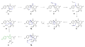
 GFP fluorophore formation.svg 512 × 83; 27 KB
-
 GFP illumination.jpg 1 553 × 1 552; 447 KB
-
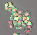
 GFP in adipocyte (2263174384).jpg 658 × 517; 16 KB
-
 GFP mechanism.svg 1 545 × 862; 335 KB
-
 GFP Superresolution Christoph Cremer.JPG 538 × 389; 156 KB
-
 Gfpantibodycell.jpg 224 × 325; 24 KB
-
 Green flourescent colonies of Escherichia coli on agar plate.png 2 142 × 1 927; 6 MB
-

-
 Green fluorescent protein image of the mouse cochlea.jpg 400 × 400; 29 KB
-
Green-to-red-photoconversion-of-GFP-for-protein-tracking-in-vivo-srep11771-s2.ogv 3,8 s, 1 032 × 1 025; 3,52 MB
-
Green-to-red-photoconversion-of-GFP-for-protein-tracking-in-vivo-srep11771-s3.ogv 17 s, 1 086 × 409; 1,13 MB
-
Green-to-red-photoconversion-of-GFP-for-protein-tracking-in-vivo-srep11771-s4.ogv 14 s, 1 032 × 1 025; 1,22 MB
-
Green-to-red-photoconversion-of-GFP-for-protein-tracking-in-vivo-srep11771-s5.ogv 22 s, 1 032 × 1 025; 944 KB
-
-
-
-
-
-
-
-
Improved-Tracking-and-Resolution-of-Bacteria-in-Holographic-Microscopy-Using-Dye-and-Fluorescent-Video1.ogv 2,4 s, 1 275 × 1 218; 1,05 MB
-
Improved-Tracking-and-Resolution-of-Bacteria-in-Holographic-Microscopy-Using-Dye-and-Fluorescent-Video10.ogv 3,3 s, 1 182 × 729; 3,05 MB
-
Improved-Tracking-and-Resolution-of-Bacteria-in-Holographic-Microscopy-Using-Dye-and-Fluorescent-Video2.ogv 2,7 s, 1 185 × 1 116; 1,37 MB
-
Improved-Tracking-and-Resolution-of-Bacteria-in-Holographic-Microscopy-Using-Dye-and-Fluorescent-Video3.ogv 2,4 s, 1 101 × 1 200; 592 KB
-
Improved-Tracking-and-Resolution-of-Bacteria-in-Holographic-Microscopy-Using-Dye-and-Fluorescent-Video4.ogv 2,7 s, 1 040 × 1 006; 637 KB
-
Improved-Tracking-and-Resolution-of-Bacteria-in-Holographic-Microscopy-Using-Dye-and-Fluorescent-Video5.ogv 3,3 s, 1 740 × 1 806; 5,6 MB
-
Improved-Tracking-and-Resolution-of-Bacteria-in-Holographic-Microscopy-Using-Dye-and-Fluorescent-Video6.ogv 3,3 s, 1 722 × 1 836; 5,35 MB
-
-
Improved-Tracking-and-Resolution-of-Bacteria-in-Holographic-Microscopy-Using-Dye-and-Fluorescent-Video8.ogv 6,7 s, 1 068 × 744; 4,58 MB
-
Improved-Tracking-and-Resolution-of-Bacteria-in-Holographic-Microscopy-Using-Dye-and-Fluorescent-Video9.ogv 3,3 s, 1 209 × 900; 3,9 MB
-
-
-
-
In-vivo-single-molecule-imaging-identifies-altered-dynamics-of-calcium-channels-in-dystrophin-ncomms5974-s5.ogv 3,9 s, 1 024 × 512; 6,83 MB
-
-
In-vivo-single-molecule-imaging-identifies-altered-dynamics-of-calcium-channels-in-dystrophin-ncomms5974-s7.ogv 9,5 s, 768 × 1 024; 48,12 MB
-
Inside-out-Ca2+-signalling-prompted-by-STIM1-conformational-switch-ncomms8826-s2.ogv 5,3 s, 1 160 × 448; 559 KB
-
Inside-out-Ca2+-signalling-prompted-by-STIM1-conformational-switch-ncomms8826-s3.ogv 5,0 s, 752 × 564; 208 KB
-
Inside-out-Ca2+-signalling-prompted-by-STIM1-conformational-switch-ncomms8826-s4.ogv 5,0 s, 1 004 × 504; 288 KB
-
Inside-out-Ca2+-signalling-prompted-by-STIM1-conformational-switch-ncomms8826-s5.ogv 6,0 s, 1 176 × 592; 89 KB
-
-
-
-
Large-scale-production-of-megakaryocytes-from-human-pluripotent-stem-cells-by-chemically-defined-ncomms11208-s5.ogv 35 s, 1 440 × 1 080; 1,29 MB
-
-
-
-
-
-
-
-
Live-cell-imaging-of-actin-dynamics-reveals-mechanisms-of-stereocilia-length-regulation-in-the-ncomms7873-s2.ogv 42 s, 2 046 × 1 730; 26,26 MB
-
Live-cell-imaging-of-actin-dynamics-reveals-mechanisms-of-stereocilia-length-regulation-in-the-ncomms7873-s3.ogv 47 s, 1 365 × 1 730; 28,71 MB
-
Live-cell-imaging-of-actin-dynamics-reveals-mechanisms-of-stereocilia-length-regulation-in-the-ncomms7873-s4.ogv 47 s, 1 364 × 1 730; 25,99 MB
-
Live-cell-imaging-of-actin-dynamics-reveals-mechanisms-of-stereocilia-length-regulation-in-the-ncomms7873-s5.ogv 36 s, 1 021 × 1 880; 37,3 MB
-
Live-cell-imaging-of-actin-dynamics-reveals-mechanisms-of-stereocilia-length-regulation-in-the-ncomms7873-s6.ogv 37 s, 1 023 × 1 384; 14,11 MB
-
Live-cell-imaging-of-actin-dynamics-reveals-mechanisms-of-stereocilia-length-regulation-in-the-ncomms7873-s7.ogv 35 s, 1 100 × 1 429; 21,05 MB
-
-
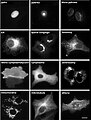
 Localisations02eng.jpg 469 × 618; 72 KB
-
 Lokalizacje.JPG 607 × 798; 57 KB
-
-
-
Long-tip-high-speed-atomic-force-microscopy-for-nanometer-scale-imaging-in-live-cells-srep08724-s2.ogv 5,6 s, 642 × 320; 1 013 KB
-
-
Long-tip-high-speed-atomic-force-microscopy-for-nanometer-scale-imaging-in-live-cells-srep08724-s4.ogv 4,5 s, 642 × 320; 1,19 MB
-
Long-tip-high-speed-atomic-force-microscopy-for-nanometer-scale-imaging-in-live-cells-srep08724-s5.ogv 6,0 s, 642 × 320; 1,36 MB
-
-
-
Long-tip-high-speed-atomic-force-microscopy-for-nanometer-scale-imaging-in-live-cells-srep08724-s9.ogv 14 s, 320 × 320; 1 005 KB
-
-
-
Mapping-the-dynamics-and-nanoscale-organization-of-synaptic-adhesion-proteins-using-monomeric-ncomms10773-s4.ogv 21 s, 1 200 × 900; 31,54 MB
-
Mapping-the-dynamics-and-nanoscale-organization-of-synaptic-adhesion-proteins-using-monomeric-ncomms10773-s5.ogv 21 s, 1 158 × 897; 25,79 MB
-
Mapping-the-dynamics-and-nanoscale-organization-of-synaptic-adhesion-proteins-using-monomeric-ncomms10773-s6.ogv 6,3 s, 1 140 × 1 140; 6,88 MB
-
-
-
-
-
-
-
-
-
-
-
-
-
-
-
Mouse-Pancreas-Tissue-Slice-Culture-Facilitates-Long-Term-Studies-of-Exocrine-and-Endocrine-Cell-pone.0078706.s002.ogv 1 min 0 s, 429 × 422; 9,25 MB
-
-
-
-
-
-
-
-
-
-
-
-
-
-
-
PEA-CLARITY-3D-molecular-imaging-of-whole-plant-organs-srep13492-s1.ogv 6,7 s, 1 280 × 720; 8,92 MB
-

-
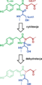 Powstawanie fluoroforu GFP.png 704 × 1 411; 31 KB
-
-
-
Probing-protein-interactions-in-living-mammalian-cells-on-a-microtubule-bench-srep17304-s2.ogv 1,5 s, 636 × 456; 238 KB
-
Probing-protein-interactions-in-living-mammalian-cells-on-a-microtubule-bench-srep17304-s3.ogv 1,2 s, 1 022 × 474; 322 KB
-
Probing-protein-interactions-in-living-mammalian-cells-on-a-microtubule-bench-srep17304-s4.ogv 2,3 s, 534 × 420; 153 KB
-
-
-
-
 S cerevisiae septins.jpg 471 × 356; 51 KB
-
 Shear Number GFP.svg 738 × 592; 114 KB
-
 Single YFP molecule superresolution microscopy-ru.png 340 × 686; 91 KB


.jpg)


.jpg)


