Category:Larvae
Jump to navigation
Jump to search
juvenile form of distinct animals before metamorphosis .jpg) Larfa buwch gota | |||||
| Upload media | |||||
| Instance of |
| ||||
|---|---|---|---|---|---|
| Subclass of |
| ||||
| Facet of |
| ||||
| Different from | |||||
| |||||
Subcategories
This category has the following 21 subcategories, out of 21 total.
A
- Acarine larvae (19 F)
- Actinotrocha (2 F)
C
E
- Ephyra (6 F)
F
G
- Glochidia (27 F)
I
M
- Metanauplii (2 F)
- Metatrochophore (5 F)
N
O
- Oyster spat (102 F)
P
- Phyllosoma (19 F)
S
T
- Trochophore (15 F)
V
- Veliger (36 F)
Z
- Zoeae (51 F)
Media in category "Larvae"
The following 200 files are in this category, out of 309 total.
(previous page) (next page)-
A-Motor-Function-for-the-DEAD-Box-RNA-Helicase-Gemin3-in-Drosophila-pgen.1000265.s002.ogv 20 s, 800 × 400; 967 KB
-
A-Motor-Function-for-the-DEAD-Box-RNA-Helicase-Gemin3-in-Drosophila-pgen.1000265.s003.ogv 8.9 s, 800 × 400; 397 KB
-
A-role-for-p38-MAPK-in-the-regulation-of-ciliary-motion-in-a-eukaryote-1471-2121-12-6-S1.ogv 25 s, 510 × 383; 1.5 MB
-
 Agua acumulada producto a fuertes lluvias.jpg 1,200 × 1,600; 350 KB
-
-
 Antlion larva - Flickr - Jay Sturner.jpg 800 × 600; 711 KB
-
 Arthropod periblastula larva.jpg 5,038 × 5,117; 3.57 MB
-
 Ascidian larvae.jpg 800 × 598; 73 KB
-
 Asota Caricae.jpg 2,608 × 2,608; 1.5 MB
-
 Auricularia larva.jpg 1,542 × 1,542; 441 KB
-
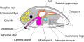 Barnacles Cypris anatomy diagram vecorized.svg 1,092 × 551; 44 KB
-
 Bivalvia gen. sp., larva 1.jpg 900 × 700; 225 KB
-
 Bivalvia gen. sp., larva 2.jpg 1,000 × 900; 320 KB
-
 Brooded coral larvae.JPG 3,648 × 2,048; 1.27 MB
-
 Bucolus fourneti late instar larvae.jpg 2,848 × 2,134; 926 KB
-
 Butterfly larva 02.JPG 4,000 × 3,000; 5.17 MB
-
 Camera 24b.jpg 576 × 562; 84 KB
-
 Camponotus ant attending larvae in Sanjay Van.jpg 4,896 × 2,752; 6.37 MB
-
 Candelabrum cocksii f.JPG 1,091 × 1,363; 174 KB
-
 Candelabrum cocksii g.JPG 1,456 × 1,096; 167 KB
-
 Cobreiros ou bicho preto.jpg 4,128 × 3,096; 5.13 MB
-
 Common Nawab Larvae.jpg 1,304 × 436; 56 KB
-
 Compound microscope used to examine meat, France, 1851-1900 Wellcome L0057251.jpg 2,832 × 4,256; 1.46 MB
-
 Coral Larvae.jpg 3,648 × 2,048; 1.43 MB
-
 Crepidula mollusk larva, or the veliger, completed.jpg 834 × 962; 350 KB
-
 De metamorphosi eleutheratorum observationes (Tab. IX) (8244679488).jpg 1,266 × 2,021; 352 KB
-
 De metamorphosi eleutheratorum observationes (Tab. VII) (8243610903).jpg 1,266 × 2,021; 336 KB
-
 De metamorphosi eleutheratorum observationes (Tab. VIII) (8243611209).jpg 1,266 × 2,021; 376 KB
-
 De metamorphosi eleutheratorum observationes (Tab. X) (8244679900).jpg 1,266 × 2,021; 338 KB
-
 De metamorphosi eleutheratorum observationes (Tab. XI) (8243612249).jpg 1,266 × 2,021; 396 KB
-
 De metamorphosi eleutheratorum observationes (Tab. XII) (8244680540).jpg 1,266 × 2,021; 349 KB
-
 De metamorphosi eleutheratorum observationes (Tab. XIV) (8244680812).jpg 1,266 × 2,021; 406 KB
-
 De metamorphosi eleutheratorum observationes (Tab. XV) (8244681084).jpg 1,266 × 2,021; 399 KB
-
 De metamorphosi eleutheratorum observationes (Tab. XVI) (8243613547).jpg 1,266 × 2,021; 395 KB
-
 De metamorphosi eleutheratorum observationes (Tab. XVII) (8244681798).jpg 1,266 × 2,021; 409 KB
-
 De metamorphosi eleutheratorum observationes (Tab. XVIII) (8243614477).jpg 1,266 × 2,021; 351 KB
-
 De metamorphosi eleutheratorum observationes (Tab. XVIII) (8243614847).jpg 1,266 × 2,021; 392 KB
-
Dermatobia hominis em movimento.webm 18 s, 640 × 352; 2.36 MB
-
 Detail of larvae damage (8716936095).jpg 3,872 × 2,592; 4.35 MB
-
 Diaphonis neri.jpg 4,608 × 3,456; 2.48 MB
-
 Dragonfly larva skin SEM stereo 70x.png 1,244 × 1,016; 2.13 MB
-
 Driopa phoebus larvae.jpg 2,592 × 1,944; 1.42 MB
-
Dtorsin-the-Drosophila-Ortholog-of-the-Early-Onset-Dystonia-TOR1A-(DYT1)-Plays-a-Novel-Role-in-pone.0026183.s006.ogv 1 min 1 s, 352 × 288; 761 KB
-
 E. amygdali- larva in situ.JPG 2,816 × 2,112; 2.34 MB
-
 E. amygdali- nymphae.jpg 1,068 × 1,293; 910 KB
-
 Echinopluteus larva.jpg 2,272 × 1,704; 375 KB
-
 Eggs and larvae of Strongyloides stercoralis.jpg 1,920 × 1,080; 191 KB
-
 Empty sheath of L3 larvae.jpg 309 × 231; 11 KB
-
 Ephemer 1.jpg 4,160 × 3,120; 2.94 MB
-
 Euproctus platycephalus larva.jpg 491 × 280; 37 KB
-
Field-Studies-Reveal-Strong-Postmating-Isolation-between-Ecologically-Divergent-Butterfly-pbio.1000529.s015.ogv 1 min 28 s, 480 × 360; 13.4 MB
-
-
Fin-Tail-Coordination-during-Escape-and-Predatory-Behavior-in-Larval-Zebrafish-pone.0032295.s002.ogv 5.6 s, 258 × 227; 629 KB
-
Fin-Tail-Coordination-during-Escape-and-Predatory-Behavior-in-Larval-Zebrafish-pone.0032295.s003.ogv 7.7 s, 224 × 133; 521 KB
-
Fin-Tail-Coordination-during-Escape-and-Predatory-Behavior-in-Larval-Zebrafish-pone.0032295.s004.ogv 7.8 s, 268 × 188; 578 KB
-
 First Larval Stage P Kadiakensis.jpg 1,720 × 5,768; 1.04 MB
-
 Fliegenlarven an Karde.jpg 2,048 × 1,536; 366 KB
-
 FMIB 48266 Egg--Embryo--Larva--To Youngest Spat.jpeg 1,057 × 1,623; 223 KB
-

-
 FMIB 49259 Forms of Surface Life- Young worm.jpeg 201 × 281; 13 KB
-
 Fmicb-14-1114849-g001.jpg 1,477 × 1,915; 432 KB
-
 Frontal view of a pluteus larva.tif 2,076 × 1,557; 7.32 MB
-
Genome-wide-analysis-of-gene-expression-during-Xenopus-tropicalis-tadpole-tail-regeneration-1471-213X-11-70-S2.ogv 1 min 12 s, 256 × 256; 1.19 MB
-
-
-
Glial-Processes-at-the-Drosophila-Larval-Neuromuscular-Junction-Match-Synaptic-Growth-pone.0037876.s005.ogv 3.0 s, 1,152 × 1,188; 471 KB
-
Glial-Processes-at-the-Drosophila-Larval-Neuromuscular-Junction-Match-Synaptic-Growth-pone.0037876.s006.ogv 1 min 10 s, 512 × 512; 230 KB
-
Glial-Processes-at-the-Drosophila-Larval-Neuromuscular-Junction-Match-Synaptic-Growth-pone.0037876.s008.ogv 2.4 s, 1,015 × 1,015; 537 KB
-
 Gombanyű.JPG 1,600 × 1,200; 303 KB
-
-
 Haeckel Asteridea Larvae Adjusted.jpg 780 × 1,278; 471 KB
-
 Haeckel Thoracostraca.jpg 2,342 × 3,289; 1.54 MB
-
 Hoechst 33342 Stain - Platynereis dumerilii larvae.jpg 1,116 × 837; 215 KB
-

-

-

-

-

-

-
-

-

-

-

-

-

-

-

-

-
-

-

-

-

-

-

-

-

-

-

-

-

-

-

-

-

-

-

-

-
 In vitro L3 Exsheathment.jpg 313 × 235; 11 KB
-
-
-
-
-
-
-
-
-
-
-
-
-
-
-
 Infective L3 Larva.jpg 313 × 233; 10 KB
-
Interspecific-Nematode-Signals-Regulate-Dispersal-Behavior-pone.0038735.s001.ogv 20 s, 680 × 512; 129 KB
-
Interspecific-Nematode-Signals-Regulate-Dispersal-Behavior-pone.0038735.s002.ogv 9.8 s, 680 × 512; 161 KB
-
Interspecific-Nematode-Signals-Regulate-Dispersal-Behavior-pone.0038735.s003.ogv 9.8 s, 680 × 512; 861 KB
-
 Inver.jpg 1,024 × 768; 559 KB
-
 Invicta hatching.tif 1,024 × 768; 769 KB
-
 Ipomoea batatas, Lochfrass, Josef Schlaghecken.jpg 4,608 × 3,456; 4.43 MB
-
 JfBoholProvinceHillsfvf 16.jpg 528 × 720; 69 KB
-
 JfLoboc7023TarsierConservationfvf 06.JPG 2,592 × 1,944; 864 KB
-
 Keket larva.jpg 6,000 × 4,000; 5.9 MB
-
 Kraliçe arı.jpg 800 × 1,200; 191 KB
-
 Lacewing larvae and egg cases (Neuroptera, F. Nymphidae) (4363852691).jpg 1,500 × 1,004; 1.82 MB
-
 Lampirys Noctiluca larva.jpg 453 × 981; 78 KB
-
 Larv (6070249639).jpg 2,272 × 1,704; 1.87 MB
-
 Larv (6070796446).jpg 2,272 × 1,704; 1.83 MB
-
 Larva (8071670256).jpg 768 × 1,024; 244 KB
-
 Larva - Algonquin Provincial Park 2019-09-24 (01).jpg 1,485 × 1,856; 1.62 MB
-
 Larva - Algonquin Provincial Park 2019-09-24 (02).jpg 1,659 × 2,074; 1.95 MB
-
 Larva - Algonquin Provincial Park 2019-09-24 (03).jpg 1,226 × 1,839; 1.37 MB
-
 Larva ascidia-ES.svg 665 × 438; 25 KB
-
 Larva ascidia-key.svg 564 × 301; 29 KB
-
 Larva de Scarabaeidae.jpg 3,747 × 3,747; 4.22 MB
-
 Larva in Water.jpg 4,160 × 3,120; 2.62 MB
-
 Larva megalopa camarao carideo.jpg 797 × 1,063; 102 KB
-
 Larva of a tunicate.jpg 400 × 261; 33 KB
-
 Larva on human hand.JPG 3,648 × 2,736; 1.89 MB
-
 Larva on Radish Stalk.jpg 720 × 960; 88 KB
-
 Larva pakomára vyskytující se v Hromnickém jezírku. .jpg 3,808 × 2,856; 880 KB
-
 Larva-Nurnberg-Germany.jpg 750 × 1,000; 280 KB
-
 Larvae evolved into chili pepper, Ларви во чили црвен пипер.JPG 4,608 × 3,300; 633 KB
-
 Larvae of coco palm butterfly (4290349577).jpg 669 × 983; 113 KB
-
 LARVAE.jpg 9,000 × 12,000; 5.66 MB
-
 Larval White Seabass (3 days post hatching) .tif 6,000 × 4,000; 68.68 MB
-
-
 Larve (Rhagium sycophanta).jpg 3,240 × 1,750; 370 KB
-
 Larve d'éphémère.JPG 477 × 377; 105 KB
-
 Larve de Scarabée rhinoceros.jpg 2,280 × 2,208; 1.56 MB
-
 Larve des Apfelwickler.jpg 2,448 × 3,264; 1.95 MB
-
 Larve di zanzare, Alpe Loasa.jpg 3,024 × 4,032; 2.05 MB
-
 Larve1.jpg 2,931 × 2,327; 2.37 MB
-
 Larven im Holzkompost 2021-04-531.jpg 2,582 × 2,582; 508 KB
-
 Larven im Holzkompost 2021-04-534.jpg 1,730 × 2,015; 246 KB
-
 Larven im Holzkompost 2021-04-537.jpg 1,331 × 1,273; 111 KB
-
 Larven im Holzkompost 2021-04-538.jpg 3,025 × 2,292; 514 KB
-
 Larwa MK (ubt).JPG 1,581 × 853; 509 KB
-
-
 LAVA MOTH.jpg 9,000 × 12,000; 6.79 MB
-
-
-
-
-
-
-
-
-
-
-
-
-
-
-
 Macro june beetle grub.jpg 945 × 629; 73 KB
-
Microgravity-simulation-by-diamagnetic-levitation-effects-of-a-strong-gradient-magnetic-field-on-1471-2164-13-52-S2.ogv 10 min 0 s, 640 × 480; 81.26 MB
-
 Miyaz.jpg 4,208 × 3,120; 10.22 MB
-
Model-Derived-Dispersal-Pathways-from-Multiple-Source-Populations-Explain-Variability-of-pone.0035794.s001.ogv 1 min 12 s, 440 × 364; 1,003 KB
-
Model-Derived-Dispersal-Pathways-from-Multiple-Source-Populations-Explain-Variability-of-pone.0035794.s002.ogv 1 min 10 s, 440 × 364; 999 KB
-
 Muga Cocoon.jpg 4,608 × 3,456; 4.78 MB
-
-
-
 Müller's Larva of Platyhelminthes.jpg 2,349 × 2,811; 1.49 MB
-
 Nauplius cirripède.JPG 1,440 × 1,232; 327 KB
-
-
-
Neuroarchitecture-of-Peptidergic-Systems-in-the-Larval-Ventral-Ganglion-of-Drosophila-melanogaster-pone.0000695.s011.ogv 8.2 s, 1,218 × 1,000; 1.45 MB
-
-


_(8244679488).jpg)
_(8243610903).jpg)
_(8243611209).jpg)
_(8244679900).jpg)
_(8243612249).jpg)
_(8244680540).jpg)
_(8244680812).jpg)
_(8244681084).jpg)
_(8243613547).jpg)
_(8244681798).jpg)
_(8243614477).jpg)
_(8243614847).jpg)
.jpg)


_(6059160203).jpg)
_(6059160661).jpg)
_(6059708482).jpg)
_(6059708694).jpg)
_(6059710142).jpg)
_(6059709010).jpg)
_(6059161761).jpg)
_(6059709566).jpg)
_(6059162237).jpg)
_(6059162807).jpg)
_(6059710676).jpg)
_(6059710976).jpg)
_(6059163935).jpg)
_(6059164139).jpg)
_(6059713418).jpg)
_(6059711878).jpg)
_(6059712172).jpg)
_(6059712576).jpg)
_(6059165513).jpg)
_(6059166309).jpg)
_(6059713960).jpg)
_(6059714192).jpg)
_(6059714408).jpg)
_(6059714732).jpg)
_(6059716192).jpg)
_(6059167503).jpg)
_(6059167851).jpg)
_(6059715574).jpg)
_(6059715872).jpg)
_(6059169019).jpg)
_(6059169341).jpg)
_(6059169625).jpg)
_(6059169837).jpg)
_(6059170189).jpg)
_(6059170425).jpg)
_(6059170777).jpg)


_(4363852691).jpg)

.jpg)
.jpg)
.jpg)
.jpg)
.jpg)
.jpg)


.jpg)

.jpg)


.JPG)


