Category:Microscopic images relating to parasites
Jump to navigation
Jump to search
Subcategories
This category has only the following subcategory.
B
Media in category "Microscopic images relating to parasites"
The following 200 files are in this category, out of 378 total.
(previous page) (next page)-
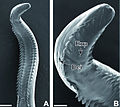
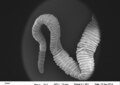 . Bothriocephalus opsariichthydis - whole parasite (27 x).tif 1,021 × 723; 723 KB
-
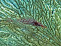 0104RajaAmpatS - 80 longnose hawkfish and parasite (5556216138).jpg 1,417 × 1,092; 276 KB
-
 04 Dilepis.tif 950 × 713; 1.96 MB
-
 06 Toxocara canis.tif 950 × 713; 1.96 MB
-
 08 Phlebotomus.tif 713 × 950; 1.96 MB
-
 12 Diclidophora denticulata.tif 637 × 950; 1.75 MB
-
 13071 2019 3727 Fig2.webp 1,595 × 869; 244 KB
-
 13071 2019 3727 Fig6.webp 1,590 × 1,851; 850 KB
-
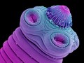
 15 Eudiplozoon nipponicum.tif 950 × 713; 1.96 MB
-
 2 trophozoite of Gregarina Garnhami.jpg 1,044 × 772; 100 KB
-
 2 trophozoites Gregarina Garnhami.jpg 1,044 × 772; 108 KB
-
 2 trophozoites2.jpg 1,044 × 772; 78 KB
-
 20 Schistosoma mansoni.tif 713 × 950; 1.96 MB
-
 2009-02-24 Taenia crassiceps.jpg 2,334 × 1,029; 1.54 MB
-
 Adult of Enterobius vermicularis.jpg 1,936 × 2,592; 1.61 MB
-
 Anterior end of Sparicotyle chrysophrii.tif 1,704 × 2,272; 11.1 MB
-
 Autofluorescence Gregarina Garnhami.jpg 904 × 299; 106 KB
-
 Balantidium coli trophozoite.jpg 727 × 567; 104 KB
-
 Bestiolina similis with Vampyrophrya pelagica infection.webp 896 × 581; 86 KB
-
 Bio 453.png 239 × 653; 148 KB
-
 Borrelia burgdorferi-cropped.jpg 346 × 395; 97 KB
-
 Chytrid parasites of marine diatoms.jpg 1,280 × 568; 105 KB
-
 CLBrenerTcruzi.jpg 194 × 251; 3 KB
-
 Clonorchis sinensis 2.png 1,122 × 382; 445 KB
-
 Collinia sp.jpg 500 × 415; 71 KB
-
 Ctenocephalides-flea-larva-2.jpg 800 × 469; 128 KB
-
 Dicrocoelium.jpg 2,313 × 3,109; 1.04 MB
-

-
 Dircrocoelimegg.jpg 2,064 × 2,652; 706 KB
-
 Egg of Enterobius vermicularis.jpg 4,000 × 2,250; 1,013 KB
-
 Egg of Faciola hepatica.jpg 3,264 × 2,448; 965 KB
-
 Egg of Hymenolepis nana.jpg 3,264 × 2,448; 936 KB
-
 Egg of Pinworm.jpg 4,000 × 2,250; 1,003 KB
-
 Egg of roundworm.jpg 3,264 × 2,448; 817 KB
-
 Egg of Sheatworm.jpg 4,000 × 2,250; 1.02 MB
-
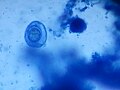
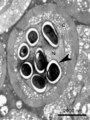
 Entaemoeba coli cyst 6 nuclei.png 180 × 178; 53 KB
-
 Entamoeba coli cysts.jpg 448 × 327; 95 KB
-
 Enteromyxum leei.jpg 1,567 × 1,752; 181 KB
-
 Enterospora nucleophila.tif 370 × 494; 69 KB
-
 Eucoleus aerophilus female vulva.jpg 945 × 1,247; 581 KB
-
 Eucoleus aerophilus in trachea of fox.jpg 945 × 630; 515 KB
-
 Exosome Secretion Affects Social Motility in Trypanosoma brucei.png 656 × 656; 164 KB
-
 Extremidad anterior a galli.jpg 970 × 735; 139 KB
-
 Figure 2 S. molnari stages.jpg 450 × 246; 12 KB
-
 Fish's parasite.jpg 2,048 × 1,536; 1.04 MB
-
 Fmars-08-720424-g002.jpg 2,367 × 2,391; 654 KB
-
 Fmars-08-720424-g003.jpg 2,367 × 1,825; 675 KB
-
 Fmars-08-720424-g004.jpg 2,367 × 1,801; 502 KB
-
 Fmars-08-742498-g002.jpg 4,563 × 3,298; 495 KB
-
 Fmars-08-742498-g003.jpg 3,162 × 4,016; 1.5 MB
-
 Four common forms of Blastocystis hominis Valzn.jpg 410 × 386; 27 KB
-
 Glycyphagus-mite-allergy-causing-2.JPG 718 × 875; 263 KB
-
 Gongylonema pulchrum nematode from man Figure 2a.jpg 1,936 × 2,584; 216 KB
-
 Gongylonema pulchrum nematode from man Figure 2b.jpg 2,448 × 2,674; 473 KB
-
 Gongylonema pulchrum nematode from man Figure 2c.jpg 375 × 514; 40 KB
-
 Gongylonema pulchrum nematode from man Full Plate 2.jpg 2,008 × 1,788; 307 KB
-
 Gregarina Garnhami.jpg 405 × 289; 100 KB
-
 H diminuta eggA.JPG 200 × 200; 15 KB
-
 H nana eggB.JPG 199 × 200; 18 KB
-
 Haemoproteus ilanpapernai parasite130095-fig1.png 2,343 × 2,740; 5.35 MB
-
 Haplozoon axiothellae.tif 629 × 897; 716 KB
-
 Haptor Monogenea Cleiodiscus.jpg 1,024 × 768; 265 KB
-
 Huevo de toascaris en campo claro.jpg 1,800 × 1,800; 951 KB
-
 Hymenolepis diminuta scolex.jpg 481 × 688; 20 KB
-
 Hymenolepis diminuta, egg - HPO.jpg 600 × 450; 20 KB
-
 Hymenolepisnana.jpg 2,893 × 2,017; 749 KB
-
 Larva of Cyathostomum catinatum - parasite of horses.jpg 527 × 460; 91 KB
-
 Larva of Triodontophorus sp. - parasite of horses 05.jpg 504 × 460; 86 KB
-
 Leishmaniavirus (dsRNA,capsid).jpg 625 × 1,257; 85 KB
-
 Micrographs of Alexandrium fundyense and Amoebophrya spp..tiff 2,098 × 2,402; 3.55 MB
-
 Moravec & Justine - Euterranova n. gen. and Neoterranova n. gen - parasite200141-fig2.png 3,484 × 4,179; 4.98 MB
-
 Moravec & Justine - Euterranova n. gen. and Neoterranova n. gen - parasite200141-fig3.png 3,484 × 4,179; 6.26 MB
-
 Moravec & Justine - Euterranova n. gen. and Neoterranova n. gen - parasite200141-fig5.png 3,484 × 2,788; 4.64 MB
-
 Moravec & Justine - Euterranova n. gen. and Neoterranova n. gen - parasite200141-fig6.png 3,484 × 3,136; 4.64 MB
-
 Moravec & Justine - New Cucullanidae - parasite200044-fig02.png 3,445 × 4,132; 5.22 MB
-
 Moravec & Justine - New Cucullanidae - parasite200044-fig03.png 3,504 × 2,804; 3.32 MB
-
 Moravec & Justine - New Cucullanidae - parasite200044-fig05.png 3,189 × 4,142; 5.17 MB
-
 Moravec & Justine - New Cucullanidae - parasite200044-fig06.png 1,581 × 2,510; 1.09 MB
-
 Moravec & Justine - New Cucullanidae - parasite200044-fig08.png 3,445 × 4,133; 4.63 MB
-
 Moravec & Justine - New Cucullanidae - parasite200044-fig10.png 3,445 × 4,133; 4.9 MB
-
 Moravec & Justine - New Cucullanidae - parasite200044-fig11.png 3,504 × 2,806; 3.42 MB
-
 Moravec & Justine - New Cucullanidae - parasite200044-fig13.png 3,189 × 4,142; 4.53 MB
-
 Moravec & Justine - New Cucullanidae - parasite200044-fig14.png 3,504 × 2,806; 3.82 MB
-
 Moravec & Justine Anisakidae 2020 parasite200028-fig02.png 2,835 × 3,402; 4.35 MB
-
 Moravec & Justine Anisakidae 2020 parasite200028-fig04.png 2,835 × 3,402; 4.56 MB
-
 Moravec & Justine Anisakidae 2020 parasite200028-fig05.png 2,835 × 2,268; 2.3 MB
-
 Moravec & Justine Anisakidae 2020 parasite200028-fig07.png 2,835 × 3,402; 3.9 MB
-
 Moravec & Justine Anisakidae 2020 parasite200028-fig08.png 1,417 × 2,268; 1.36 MB
-
 Moravec & Justine Anisakidae 2020 parasite200028-fig10.png 2,835 × 3,402; 3.94 MB
-
 Moravec & Justine Anisakidae 2020 parasite200028-fig11.png 2,835 × 3,685; 4.41 MB
-
 Moravec & Justine Anisakidae 2020 parasite200028-fig13.png 3,154 × 3,784; 4.57 MB
-
 Moravec & Justine Anisakidae 2020 parasite200028-fig14.png 2,835 × 2,268; 2.43 MB
-
 Moravec & Justine Anisakidae 2020 parasite200028-fig16.png 2,835 × 3,685; 4.67 MB
-
 Moravec & Justine Anisakidae 2020 parasite200028-fig17.png 2,835 × 2,268; 2.35 MB
-
 Moravec & Justine Spirurida 2020 parasite190153-fig2.png 2,662 × 2,131; 2.76 MB
-
 Moravec & Justine Spirurida 2020 parasite190153-fig5.png 2,662 × 3,194; 3.84 MB
-
 Moravec & Justine Spirurida 2020 parasite190153-fig6.png 2,662 × 3,459; 3.47 MB
-
 Moravec & Justine Spirurida 2020 parasite190153-fig8.png 2,657 × 3,188; 3 MB
-
 Moravec & Justine Spirurida 2020 parasite190153-fig9.png 2,662 × 2,131; 2.18 MB
-
 Necator1.jpg 947 × 818; 113 KB
-
 Necator2.jpg 4,128 × 2,322; 1.16 MB
-
 Necator3.jpg 799 × 921; 56 KB
-
 NematodePNWD USDA.jpg 300 × 230; 138 KB
-
 Paracalanid copepods infected by Ellobiopsis chattoni (horizontal).jpg 1,080 × 193; 77 KB
-
 Paracalanid copepods infected by Ellobiopsis chattoni.jpg 538 × 377; 36 KB
-
 Parasite 20, 51 (2013) Trypanosoma (Megatrypanum) lainsoni Figs 1-20.tif 2,067 × 2,900; 2.13 MB
-
 Parasite 20, 51 (2013) Trypanosoma (Megatrypanum) lainsoni Figs 1-3.jpg 1,517 × 532; 272 KB
-
 Parasite 20,42(2013) Complete life cycle of a pennellid Peniculus minuticaudae -fig2.tif 1,654 × 1,141; 753 KB
-
 Parasite 20,43 (2013) Redescription of Setaria graberi (Nematoda, Filarioidea) Figure 4.tif 1,772 × 1,743; 2.38 MB
-
 Parasite130039 Pterydodermatites quentini -fig2.jpg 2,343 × 2,398; 2.32 MB
-
 Parasite130039 Pterydodermatites quentini -fig3 Figure 3 A-B.jpg 1,243 × 1,108; 304 KB
-
 Parasite130039 Pterydodermatites quentini -fig3.tif 2,343 × 2,210; 3.3 MB
-
 Parasite130049 Haemoproteus syrnii -fig1.jpg 1,803 × 2,825; 2.53 MB
-
 Parasite130049 Haemoproteus syrnii -fig2.jpg 2,343 × 2,278; 2.45 MB
-
 Parasite130049 Haemoproteus syrnii -fig3.jpg 1,654 × 1,632; 1.69 MB
-
 Parasite130059-fig1 Spermiogenesis in Pleurogenidae (Digenea).tif 2,343 × 2,124; 1.58 MB
-
 Parasite130059-fig3 Spermiogenesis in Pleurogenidae (Digenea).tif 2,234 × 2,756; 4.64 MB
-
 Parasite130059-fig4 Spermiogenesis in Pleurogenidae (Digenea).tif 2,343 × 2,894; 5.33 MB
-
 Parasite130059-fig5 Spermiogenesis in Pleurogenidae (Digenea).tif 2,280 × 2,756; 4.48 MB
-
 Parasite130059-fig6 Spermiogenesis in Pleurogenidae (Digenea).tif 2,343 × 2,689; 4.57 MB
-
 Parasite130059-fig7 Spermiogenesis in Pleurogenidae (Digenea).tif 2,343 × 1,198; 2.61 MB
-
 Parasite130094-fig2 Cysts and laminated layer.tif 1,102 × 826; 1.22 MB
-
 Parasite130094-fig3 Cysts.tif 1,654 × 1,242; 3.51 MB
-
 Parasite130103-fig1 Protopolystoma xenopodis (Monogenea, Polystomatidae) egg.tif 2,067 × 2,079; 1.69 MB
-
 Parasite130103-fig2 Protopolystoma xenopodis (Monogenea, Polystomatidae) Oncomiracidium.tif 2,067 × 2,027; 4.81 MB
-
 Parasite130103-fig4 Protopolystoma xenopodis (Monogenea, Polystomatidae) Adult.tif 2,067 × 2,044; 2.24 MB
-

-
 Parasite130116-1-olm Entamoeba gingivalis microscopy.tif 1,436 × 1,072; 1,010 KB
-

-
-

-
 Parasite140007-fig9 Philometra selaris Moravec & Justine, 2014 (Nematoda, Philometridae).tif 2,343 × 2,811; 4.14 MB
-
 Parasite140013-fig2 Pterygodermatites (Paucipectines) baiomydis SEM.tif 2,343 × 2,257; 3.42 MB
-
 Parasite140013-fig3 Pterygodermatites (Paucipectines) baiomydis Photo.tif 1,102 × 896; 1.68 MB
-
 Parasite140015-fig1 Protoopalina pingi (Opalinidae) SEM.tif 2,343 × 1,758; 1.59 MB
-
 Parasite140015-fig2 Protoopalina pingi (Opalinidae) Microscopy.tif 2,343 × 2,896; 10.12 MB
-
 Parasite140019-fig1 Nosema podocotyloidis - Hyperparasitic Microsporidia.tif 2,022 × 3,031; 4 MB
-
 Parasite140019-fig2 Nosema podocotyloidis - Hyperparasitic Microsporidia.tif 2,453 × 2,585; 3.74 MB
-
 Parasite140019-fig3 Nosema podocotyloidis - Hyperparasitic Microsporidia.tif 2,453 × 2,204; 3.68 MB
-
 Parasite140019-fig4 Nosema podocotyloidis - Hyperparasitic Microsporidia.tif 2,453 × 2,531; 3.14 MB
-
 Parasite140027-fig2 Histological sections of Dictyocoela diporeiae.tif 1,654 × 1,240; 3.97 MB
-
-
-

-
 Parasite140065-fig1 Hemogregarines.tif 2,343 × 1,557; 5.37 MB
-
 Parasite140065-fig2 Foleyella furcata.tif 2,343 × 1,731; 6.88 MB
-
 Parasite140068-fig1 Spermatozoon of Lecithochirium microstomum (Digenea) Figs 1-6.tif 2,067 × 2,554; 3.47 MB
-
 Parasite140068-fig2 Spermatozoon of Lecithochirium microstomum (Digenea) Figs 7-12.tif 2,067 × 2,554; 3.78 MB
-
 Parasite140068-fig3 Spermatozoon of Lecithochirium microstomum (Digenea) Figs 19.tif 2,067 × 2,067; 2.89 MB
-
 Parasite140068-fig4 Spermatozoon of Lecithochirium musculus (Digenea) Figs 20-27.tif 2,067 × 2,435; 3.26 MB
-

-
 Parasite140076-fig1 Dirofilaria repens removed from a subcutaneous nodule - Photos.png 1,645 × 2,894; 5.31 MB
-

-

-

-

-
-

-
-
-
-

-

-

-

-

-

-
-

-

-

-
 Parasite140083-fig5 Figs 31-36 Cathayacanthus spinitruncatus.tif 2,067 × 2,499; 2.64 MB
-
 Parasite140083-fig6 Figs 37-44 Cathayacanthus spinitruncatus.tif 2,067 × 2,499; 3.42 MB
-
 Parasite140085-fig3 Echinococcus granulosus cysts from boiled livers and lungs of sheep.tif 2,039 × 2,998; 4.78 MB
-
 Parasite140088-fig1 The genus Tunga (Siphonaptera,Tungidae) Neosome.tif 1,654 × 747, 2 pages; 1.07 MB
-
 Parasite140088-fig2 The genus Tunga (Siphonaptera, Tungidae) A neosome of Tunga penetrans.tif 1,102 × 975, 2 pages; 2 MB
-
 Parasite140098-fig1 Collyricloides massanae sperm.tif 2,067 × 2,702; 3.69 MB
-
 Parasite140098-fig2 Collyricloides massanae sperm.tif 2,067 × 2,160; 3.01 MB
-
 Parasite140098-fig3 Collyricloides massanae sperm.tif 1,996 × 2,839; 3.79 MB
-
 Parasite140104-fig2 Surra (Trypanosoma evansi infection) in a Tunisian dog.png 1,102 × 827; 1.57 MB
-
 Parasite140105-fig1 Toxoplasmosis in a bar-shouldered dove - histology.tif 1,654 × 2,057; 3 MB
-
 Parasite140105-fig2 Toxoplasmosis in a bar-shouldered dove - histology.tif 1,378 × 1,117; 828 KB
-
 Parasite140105-fig3 Toxoplasmosis in a bar-shouldered dove - TEM of 2 tachyzoites.tif 1,378 × 1,683; 2.11 MB
-
 Parasite140120-fig3 Acanthamoeba keratitis Figure 3A.png 2,080 × 2,102; 2.38 MB
-
 Parasite140120-fig3 Acanthamoeba keratitis Figure 3B.png 2,032 × 2,102; 2.71 MB
-
 Parasite140120-fig3 Acanthamoeba keratitis.png 4,200 × 2,102; 5.19 MB
-
 Parasite140120-fig4 Acanthamoeba keratitis Figure 4A.png 1,951 × 1,985; 765 KB
-
 Parasite140120-fig4 Acanthamoeba keratitis Figure 4B.png 1,960 × 1,985; 741 KB
-
 Parasite140120-fig4 Acanthamoeba keratitis Figure 4C.png 1,911 × 1,985; 709 KB
-
 Parasite140120-fig4 Acanthamoeba keratitis.png 5,950 × 1,985; 2.2 MB
-
 Parasite140120-fig5 Acanthamoeba keratitis.png 827 × 827; 979 KB
-
 Parasite140121-fig3 Pseudorhabdosynochus jeanloui (Monogenea, Diplectanidae) Fig3.png 4,205 × 2,502; 5.18 MB
-

-

-
-
-
 Parasite140131-fig2 Capillaria plectropomi (Nematoda) - Scannin Electron Microscopy.tif 2,835 × 3,402, 2 pages; 7.15 MB
-

-
 Parasite140132-fig2 Philometra protonibeae (Nematoda, Philometridae).png 2,067 × 2,480; 3.09 MB
-
 Parasite140132-fig3 Philometra protonibeae (Nematoda, Philometridae).png 1,103 × 1,102; 1.89 MB
.jpg)


.jpg)


_in_a_Tunisian_dog.png)


_Fig3.png)
_Fig3a_Sclerotized_reproductive_organs.png)
_Fig3b_Haptor.png)
_Fig3d_Squamodisc_SEM.png)
.png)
.png)
.jpg)
_Trypanosoma_(Megatrypanum)_lainsoni_Figs_1-3.jpg)
_Fig3c_Haptor.png)