Category:Mikael Häggström/Medicine-related photographs
Jump to navigation
Jump to search

These images were created by Mikael Häggström, M.D.
- User info
- Reusing images
Subcategories
This category has only the following subcategory.
M
Media in category "Mikael Häggström/Medicine-related photographs"
The following 45 files are in this category, out of 45 total.
-
 Ancient roman folding handle for a surgical drill.jpg 2,125 × 259; 169 KB
-
 Ancient roman rectal speculum.jpg 801 × 1,461; 428 KB
-
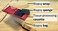 Biopsy wrap, biopsy sponge, tissue processing cassette and biopsy bag.jpg 4,681 × 2,537; 2.51 MB
-

-

-
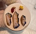
 Circle of salt and pepper.jpg 2,029 × 1,963; 1.39 MB
-
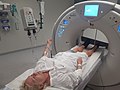 Contrast CT.jpg 3,264 × 2,448; 1.71 MB
-
 Eosin solution.jpg 2,229 × 1,721; 780 KB
-
 Fluoroscopy room with control space.jpg 1,131 × 848; 309 KB
-
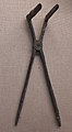
 Forceps, bone saw and bleeding cup descriptions.jpg 2,373 × 2,097; 932 KB
-
 Forceps, bone saw and bleeding cup.jpg 1,589 × 1,233; 389 KB
-
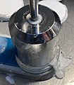
 Frozen sectioning 1 putting embedding medium on chuck.jpg 2,745 × 1,922; 1.2 MB
-
-

-
 Frozen sectioning 12 Continuing until all the tissue of interest is in the section.jpg 1,737 × 1,645; 781 KB
-
 Frozen sectioning 13 Putting a glass slide on the tissue - version 2.jpg 4,032 × 3,024; 1.53 MB
-
 Frozen sectioning 13 Putting a glass slide on the tissue.jpg 3,377 × 2,745; 2.26 MB
-
 Frozen sectioning 2 waiting until bottom is frozen.jpg 1,749 × 1,445; 548 KB
-
 Frozen sectioning 3 putting cryostat oil on conductor.jpg 1,085 × 1,181; 350 KB
-
 Frozen sectioning 4 putting conductor on chuck.jpg 1,469 × 1,421; 533 KB
-
 Frozen sectioning 5 putting specimens on chuck.jpg 1,905 × 1,793; 779 KB
-
 Frozen sectioning 6 Covering with embedding medium.jpg 1,485 × 1,333; 490 KB
-
 Frozen sectioning 7 apply conductor.jpg 1,405 × 1,605; 482 KB
-
 Frozen sectioning 8 using freeze spray.jpg 4,032 × 3,024; 3.31 MB
-
 Frozen sectioning 9 Breaking off embedding medium that reach below the chuck's plate.jpg 1,617 × 1,281; 485 KB
-
 Hematoxylin solution.jpg 2,353 × 1,805; 839 KB
-
 Leica microscopy slide scanner, annotated.jpg 3,957 × 1,584; 1.69 MB
-
 Leica microscopy slide scanner.jpg 4,032 × 3,024; 2.74 MB
-
 Micrography with a smartphone.jpg 3,705 × 2,951; 2.54 MB
-
 Microscopy slide.jpg 2,597 × 2,065; 1,014 KB
-
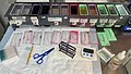
 Multitarget microbial panel (MicroScan WalkAway).jpg 4,032 × 3,024; 2.63 MB
-
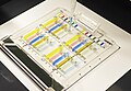
 Multitarget microbial panel for MicroScan WalkAway system.jpg 2,849 × 2,113; 1.78 MB
-
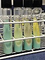
 Mycobacteria Growth Indicator Tube (MGIT) samples in ultraviolet light.jpg 4,032 × 3,024; 2.04 MB
-
 Perls Prussian blue components.jpg 3,241 × 1,909; 1.41 MB
-
 Pigtail catheter settings.jpg 2,877 × 2,549; 1.4 MB
-
 Pipetting anti-immunoglobulins to immunofixation panel.jpg 2,980 × 2,052; 1.04 MB
-
 Removing marks from a microscopy slide.jpg 1,961 × 1,037; 520 KB
-
 Right or left renal stone.jpg 1,194 × 720; 255 KB
-
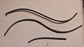 Roman catheters.jpg 1,576 × 887; 518 KB
-
 Roman spatulas.jpg 539 × 1,591; 314 KB
-
 Setup for HE staining of frozen section slides.jpg 3,953 × 2,237; 2.55 MB
-
-
 Staphylococcus aureus on blood agar, mildly backlit.jpg 4,032 × 3,024; 1.29 MB
-
 Time in solutions for frozen sections.jpg 1,500 × 1,532; 628 KB
-
 Touch prep on a lymph node.jpg 3,525 × 1,701; 1.08 MB


.jpg)

_samples_in_ultraviolet_light.jpg)


