Category:Osteoblasts
Jump to navigation
Jump to search
bone-forming cells which secrete an extracellular matrix  | |||||
| Upload media | |||||
| Instance of | |||||
|---|---|---|---|---|---|
| Subclass of |
| ||||
| Different from | |||||
| |||||
Media in category "Osteoblasts"
The following 33 files are in this category, out of 33 total.
-
 201102 osteoblast.png 417 × 566; 89 KB
-
-
 Active osteoblasts.jpg 800 × 520; 109 KB
-
Automated-measurement-of-cell-motility-and-proliferation-1471-2121-6-19-S1.ogv 38 s, 320 × 240; 2.43 MB
-
Automated-measurement-of-cell-motility-and-proliferation-1471-2121-6-19-S2.ogv 1 min 19 s, 320 × 240; 687 KB
-
Automated-measurement-of-cell-motility-and-proliferation-1471-2121-6-19-S3.ogv 1 min 51 s, 720 × 480; 1.23 MB
-
 Bone forming cells - Osteoblasts -- Smart-Servier.jpg 10,240 × 5,760; 1.27 MB
-
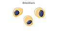
 Bone forming cells - Osteoblasts 1 -- Smart-Servier.png 1,379 × 1,318; 192 KB
-
 Bone forming cells - Osteoblasts 2 -- Smart-Servier.png 1,465 × 1,445; 200 KB
-
 Bone forming cells - Osteoblasts 3 -- Smart-Servier.png 1,252 × 1,453; 188 KB
-
 HDI effect on bone remodeling.png 3,000 × 1,099; 200 KB
-
 Kolm osteoblasti meditsiinilise titaani pinnal.jpg 1,280 × 960; 753 KB
-
-
-
-
-
-
-
 Marrow Adipocytes are derived from mesenchymal stem cell (MSC) differentiation. .tif 4,433 × 4,766; 80.6 MB
-
 Marrow Adipocytes are derived from mesenchymal stem cell (MSC) differentiation.png 4,433 × 4,766; 1.26 MB
-
 MSC diagram into osteoblast and chondrocyte cell lineages.jpg 1,148 × 624; 54 KB
-
 Osteoblast togopic.png 417 × 566; 94 KB
-
 Osteoblast.jpg 1,699 × 1,538; 968 KB
-
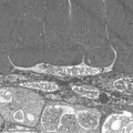
 Osteocyte.tif 2,011 × 1,441; 3.93 MB
-
-
-
Persistent-directional-cell-migration-requires-ion-transport-proteins-as-direction-sensors-and-1471-2121-12-4-S7.ogv 3.8 s, 1,376 × 1,027; 183 KB
-
-
Stochasticity-and-the-Molecular-Mechanisms-of-Induced-Pluripotency-pone.0003086.s002.ogv 10 s, 404 × 202; 130 KB
-
Stochasticity-and-the-Molecular-Mechanisms-of-Induced-Pluripotency-pone.0003086.s003.ogv 10 s, 404 × 202; 132 KB
-
Stochasticity-and-the-Molecular-Mechanisms-of-Induced-Pluripotency-pone.0003086.s004.ogv 8.0 s, 404 × 202; 112 KB
-
Stochasticity-and-the-Molecular-Mechanisms-of-Induced-Pluripotency-pone.0003086.s005.ogv 13 s, 404 × 202; 150 KB
-
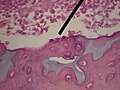 WVSOM Osteoblast.JPG 2,816 × 2,112; 2.08 MB


_differentiation.png)


