Category:Stem cells
Jump to navigation
Jump to search
Deutsch: Stammzellen
English: Stem cells
Русский: Стволовые клетки
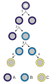
undifferentiated biological cells that can differentiate into specialized cells  | |||||
| Upload media | |||||
| Instance of | |||||
|---|---|---|---|---|---|
| Subclass of |
| ||||
| |||||
Subcategories
This category has the following 26 subcategories, out of 26 total.
?
- Induced pluripotent stem cells (175 F)
A
- Adult stem cells (87 F)
E
H
- Hematopoietic stem cells (46 F)
I
L
- Limbal stem cell (2 F)
M
- Multipotent stem cells (9 F)
N
- Neural stem cells (147 F)
O
- Oogonial stem cells (1 F)
P
- Precursor cells (19 F)
- Progenitor cells (8 F)
S
- Stem cell factor (7 F)
- Stem cell lines (21 F)
- Stem cell niche (63 F)
- Stem cell transplantation (34 F)
- Stem-cell therapy (33 F)
T
- Tumor stem cell assay (5 F)
X
- Xenobots (7 F)
Media in category "Stem cells"
The following 174 files are in this category, out of 174 total.
-
 1-s2.0-S1934590923001340-gr2 lrg.jpg 1,294 × 891; 517 KB
-
 10x-pad7-3-center m.tif 512 × 512; 446 KB
-
 422 Feature Stem Cell new.png 1,336 × 1,523; 526 KB
-
 422 Feature Stem Cell.jpg 1,005 × 1,146; 354 KB
-
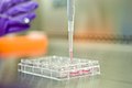 A cell culture plate with induced pluripotent stem cells (46291373491).jpg 5,416 × 3,611; 2.19 MB
-
 Alexander A. Maximow 1909.jpg 2,507 × 1,871; 585 KB
-
 Antonio e Biagio e Cesare Arrigo Cryopreservation Stem.jpg 3,264 × 2,448; 3.83 MB
-
 Antonio e Biagio e Cesare Arrigo Cryopreservation.jpg 4,608 × 3,456; 2.85 MB
-
 Antonio e Biagio e Cesare Arrigo Stem.jpg 4,608 × 3,456; 4.19 MB
-
 AplicacionesCélulas iPS-GL.jpg 960 × 720; 103 KB
-
 AplicacionesCélulas iPS.jpg 960 × 720; 70 KB
-
 Bangkokstemcell.jpg 2,048 × 1,152; 444 KB
-
 BioTek-Wikipedia-Image.tif 852 × 630; 2.6 MB
-
 BMDM production.png 720 × 405; 39 KB
-
 Bone-marrow-publ.1868.jpg 521 × 800; 270 KB
-
 Brinster Male germline stem cell transplantation.tif 2,700 × 2,007; 15.53 MB
-
-
 Cellules souches embryonnaires HD90.jpg 2,048 × 1,536; 623 KB
-
 Cellules souches embryonnaires HD90.tif 2,048 × 1,536; 6 MB
-
 Cerebral organoid flowchart.png 401 × 683; 57 KB
-
 Classification of the different types of connective tissues.png 1,448 × 700; 708 KB
-
 CSIRO ScienceImage 11406 Using a microscope to examine a slide.jpg 1,771 × 2,657; 2.94 MB
-
 CSIRO ScienceImage 1521 iPS cells in the lab.jpg 5,184 × 3,456; 5.41 MB
-
 Células madre meristemáticas 1.jpg 649 × 557; 46 KB
-
 Células Madre para Cirugías de Menisco.png 960 × 960; 332 KB
-
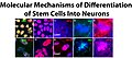 Differentiation of Stem Cells Into Neurons.jpg 1,884 × 835; 845 KB
-
 Doctor-performing-research-with-stem-cells.jpg 3,716 × 5,574; 9.17 MB
-
 Doctor-preparing-to-perform-stem-cell-therapy.jpg 3,213 × 4,820; 7.05 MB
-
 Dr. Eugene Zadorin develops a treatment protocol for a patient with liver cirrhosis.jpg 1,066 × 1,600; 173 KB
-
 Dr. Eugene Zadorin develops a treatment protocol for a patient.jpg 1,066 × 1,600; 237 KB
-
 Electron micrograph of a Fractone.jpg 3,662 × 3,472; 2.37 MB
-
 Embryonal cardiac stem cellתמונה2.jpg 505 × 455; 45 KB
-
 Embryonic origins of skeletal muscles mouse embryo.png 1,573 × 695; 425 KB
-
 Entnahme von erkrankten Stammzellen.jpg 1,632 × 920; 395 KB
-
 Erythropoietin and formation of red blood cells.JPG 2,118 × 2,480; 1.98 MB
-
 ESEM Leptospermum.svg 3,721 × 1,950; 433 KB
-
 FatStemCells.gif 260 × 208; 29 KB
-
 FatStemCellsPic.gif 252 × 100; 27 KB
-
 Final stem cell differentiation (1).svg 512 × 362; 261 KB
-
 Final stem cell differentiation es.svg 512 × 362; 277 KB
-
 Final stem cell differentiation fi.svg 512 × 362; 277 KB
-
 Final stem cell differentiation is.svg 512 × 362; 241 KB
-
 Frank Peralta.png 1,500 × 1,134; 131 KB
-
 From mesenchymal stem cells to specific connective tissue cell types.png 1,085 × 877; 337 KB
-
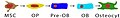 From MSC to Osteocyte.jpg 1,000 × 150; 52 KB
-
 From muscle progenitors to muscle fibers.png 1,230 × 414; 93 KB
-
 GFPPupsAndTestisForWiki.jpg 1,200 × 1,704; 627 KB
-
 Hematopoietic stem cell.jpg 1,000 × 1,500; 273 KB
-
 Hiire embrüonaalsed tüvirakud diferentseerumas neuraalseteks eelasrakkudeks.JPG 2,560 × 1,920; 2.72 MB
-
 HMSC MAP4GFP H2BRFP.jpg 518 × 514; 32 KB
-
 Human blastoid - 1.jpg 2,835 × 2,835; 2.42 MB
-
 Human blastoid.jpg 1,089 × 1,089; 1.24 MB
-
 Human bone marrow derived MSCs.jpg 1,360 × 1,024; 199 KB
-
 Human Brain Organoid.jpg 1,940 × 1,930; 464 KB
-
 Human Cell Groups distributed by Cell Count and by Aggregate Cell Mass.jpg 3,162 × 2,096; 1.08 MB
-
 Human Cell Groups; Cell Count, Cell Mass, and Aggregate Cell Mass (Biomass).png 1,258 × 847; 149 KB
-
 Human embryonic stem cell colony phase.jpg 1,024 × 894; 255 KB
-
 Human embryonic stem cells only A.png 1,010 × 752; 1.75 MB
-
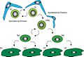 Illustration of stem cell control by the niche..jpg 550 × 369; 78 KB
-
 Induced pluripotent stem cellתמונה1.jpg 407 × 431; 43 KB
-
 Induction of iPS cells.svg 840 × 420; 12 KB
-
-
 Intestinal organoid.PNG 1,148 × 1,106; 2.4 MB
-
 Ips cells ja.png 2,583 × 2,333; 373 KB
-
 Ips cells-es.png 2,022 × 1,928; 816 KB
-
 Ips cells.png 647 × 617; 69 KB
-
 IPS process.png 773 × 521; 81 KB
-
 J. David Gladstone Institutes (4837355699).jpg 397 × 266; 21 KB
-
 Kelvin Gonell.png 905 × 960; 864 KB
-

-
 KM Transplantat.JPEG 480 × 640; 43 KB
-
 Limbal epithelium. Cells. SEM-BSE.jpg 3,072 × 2,304; 4.31 MB
-

-
 LT-HSCs are in the quiescent state..jpg 600 × 580; 87 KB
-
 Many human blastoids.jpg 1,500 × 671; 620 KB
-
-

-
 Mapping SHIV infection in the body, 2018 - Wellcome Photography Prize 2019.jpg 1,920 × 1,920; 512 KB
-
 Mesenchymal stem cell.png 610 × 210; 69 KB
-

-
 Microscopic image of stem cells, Hues 9 stained with DAPI (blue).jpg 640 × 640; 18 KB
-
 Microscopic image of stem cells, Hues 9 stained with DAPI.png 1,360 × 1,024; 1.9 MB
-

-
 Mini beating hearts N Zhelev.jpg 3,493 × 2,880; 5.29 MB
-
 MLP to DN3 resized annotated-Aug 3 2010.jpg 984 × 656; 196 KB
-
 Modelul ciclului de viata atavistic al cancerului si "familia" de celule stem.jpg 2,480 × 3,502; 793 KB
-
 MSC high magnification.jpg 1,803 × 2,400; 1.67 MB
-
 Myometrial stem cells analyses..jpg 500 × 594; 123 KB
-
 Neuron in tissue culture.jpg 1,315 × 1,033; 410 KB
-
 No chimera SSCs.jpg 720 × 540; 809 KB
-
 Pan sb.jpg 100 × 111; 2 KB
-
 Parkinson's induced pluripotent stem cell.jpg 1,800 × 1,813; 250 KB
-
 Picture1g.png 297 × 185; 78 KB
-
 Platelets by budding off from megakaryocytes.jpg 1,058 × 794; 80 KB
-
 Potencia-gl.jpg 960 × 720; 80 KB
-
 Production of iPSC Timeline.png 1,065 × 360; 316 KB
-
 Regeneração – Epimorfose e Morfalaxia.jpg 1,280 × 1,220; 252 KB
-

-
 Research-with-umbilical-cord-mesenchymal-stem-cells.jpg 4,000 × 6,000; 11.2 MB
-
 Role of Autophagy and primary cilia in embryonic stem cell lineage specification.png 3,128 × 2,729; 815 KB
-
 Role of primary cilia in Wntβ-catenin regulation and stem cell biology.png 3,049 × 2,523; 948 KB
-

-
 RUH PMH.jpg 2,766 × 3,200; 1.33 MB
-
 Schema migratie stamcellen.jpg 960 × 720; 50 KB
-

-
 Schematic overview view of niche components..jpg 350 × 139; 24 KB
-

-

-
 Schematic representation of the use of BMSCs as vectors..jpg 700 × 550; 77 KB
-

-

-

-
 Sequential soluble factors specify differentiation programs..jpg 381 × 143; 15 KB
-
 Sources of new adult β-cells..jpg 600 × 329; 86 KB
-
 STAP,iPS.png 950 × 1,140; 131 KB
-
 Stem cell differentiation.svg 512 × 362; 250 KB
-
 Stem cell distribution in the cnidarian Hydra vulgaris.png 2,616 × 4,938; 12.32 MB
-
 Stem cell division and differentiation.svg 200 × 400; 36 KB
-
 Stem Cell Research (0436) (13982409504).jpg 2,784 × 1,848; 2.15 MB
-
 Stem Cell Research (0443) (13981985775).jpg 1,725 × 2,598; 1.85 MB
-
 Stem Cell Research (0449) (14001969013).jpg 1,770 × 2,666; 1.9 MB
-
 Stem Cell Research (0456) (13978787731).jpg 2,784 × 1,848; 2.63 MB
-
 Stem Cell Research (0504) (13982408424).jpg 1,636 × 2,338; 2.04 MB
-
 Stem Cell Research.jpg 2,784 × 1,848; 2.38 MB
-
 Stem cell togopic.png 500 × 375; 88 KB
-
 Stem cell treatment.jpg 600 × 600; 53 KB
-
 Stem cell treatments.png 2,417 × 1,983; 1.38 MB
-
 Stem cell treatments.svg 1,450 × 1,190; 427 KB
-
 Stem Cell.jpg 848 × 545; 84 KB
-
 Stem cell2 togopic.png 260 × 291; 70 KB
-
 Stem Cell2.png 533 × 311; 136 KB
-
 Stem Cells (50160258728).jpg 1,157 × 1,027; 107 KB
-
 Stem cells diagram EST.png 1,015 × 927; 195 KB
-
 Stem cells diagram nl.png 1,015 × 927; 218 KB
-
 Stem cells diagram russia.png 1,015 × 927; 198 KB
-
 Stem cells diagram uk.png 1,015 × 927; 179 KB
-
 Stem cells diagram.png 1,015 × 927; 170 KB
-
 Stem cells division.svg 744 × 1,169; 11 KB
-
 Stem cells, day 1 after passage.tiff 1,392 × 1,040; 4.14 MB
-
 Stem cells, day 3 after passage.jpg 1,388 × 1,040; 1.11 MB
-
 Stem cells, day 3 after passage.tiff 1,392 × 1,040; 4.14 MB
-
 Stem cells, Hues 9 p 23.jpg 1,388 × 1,040; 660 KB
-
 Stem cells, Hues 9 p 38, day 1 after passage.jpg 1,384 × 1,036; 830 KB
-
 Stem cells, Hues 9 p 38, day 2 after passage.jpg 1,384 × 1,036; 1.01 MB
-
 Stem cells, Hues 9 stained.jpg 640 × 640; 23 KB
-
 Stem cells, Hues 9, day 2 after passage.tiff 1,392 × 1,040; 4.14 MB
-
 Stem Cells.TIF 1,030 × 1,030; 3.04 MB
-
 Stem cells2.png 640 × 1,115; 133 KB
-
 Stem-cell-therapy-for-multiple-sclerosis-1.jpg 3,949 × 5,924; 11.74 MB
-
 Stem-cell-therapy-for-multiple-sclerosis-2.jpg 3,821 × 5,731; 10.39 MB
-
 Stem-cells-being-drawn-up-for-research.jpg 3,065 × 4,597; 7.66 MB
-
 Stemcell application.tif 1,146 × 1,146; 210 KB
-
 Stimulation of c-Met leads to enhanced cancer stem cell (CSC) properties.jpg 1,772 × 822; 215 KB
-

-

-
 Telocytes-Fig 13.tif 993 × 1,180; 2.17 MB
-
 Telocytes-Fig 15.tif 1,604 × 1,792; 5.64 MB
-

-
 The development and the ways to rejuvenate cells - en.svg 2,339 × 1,629; 871 KB
-
 The development and the ways to rejuvenate cells - ru.svg 2,339 × 1,629; 881 KB
-

-
-
 Time-lapse video of dividing cells.gif 280 × 182; 7.57 MB
-
 Tissue Engineering.png 2,250 × 2,100; 1.01 MB
-
 Tumor-en.svg 522 × 782; 121 KB
-
 Tumor-nl.svg 522 × 782; 130 KB
-
 Umbilical-cord-stem-cells-in-vial.jpg 6,000 × 4,000; 11.25 MB
-
 Vial cultivated stem cells.jpg 3,264 × 2,448; 1.45 MB
-
 Vials (32419676518).jpg 5,760 × 3,840; 3.42 MB
-
 VidaCord imagenes.jpg 400 × 400; 143 KB
-
 Whartons-jelly-mesenchymal-stem-cells.jpg 4,000 × 6,000; 11.63 MB
-
 Working with induced pluripotent stem cells (46291373691).jpg 5,545 × 3,697; 2.57 MB
-
 Πολλαπλασιασμός νευρικών βλαστικών κυττάρων ποντικών.jpg 924 × 836; 113 KB
-


.jpg)


.svg)


.png)


.jpg)


.jpg)
.jpg)


.jpg)

_and_DAPI_(blue).jpg)


..jpg)


_and_promising_future_strategies_(C_and_D)_to_stimulate_anti_tumor_immune_responses_(in_orange)..jpg)


,_flow_perfusion_of_0.jpg)


_(13982409504).jpg)
_(13981985775).jpg)
_(14001969013).jpg)
_(13978787731).jpg)
_(13982408424).jpg)


.jpg)


_properties.jpg)


_and_the_subgranular_zone.jpg)


.jpg)


.jpg)

