File:Computed tomography of human brain - large.png

元のファイル (3,639 × 2,595 ピクセル、ファイルサイズ: 3.9メガバイト、MIME タイプ: image/png)
キャプション
キャプション
| このファイルはクリエイティブ・コモンズ CC0 1.0 全世界 パブリック・ドメイン提供のもとで利用可能にされています。 | |
| ある作品に本コモンズ証を関連づけた者は、その作品について世界全地域において著作権法上認められる、その者が持つすべての権利(その作品に関する権利や隣接する権利を含む。)を、法令上認められる最大限の範囲で放棄して、パブリック・ドメインに提供しています。
この作品は、たとえ営利目的であっても、許可を得ずに複製、改変・翻案、配布、上演・演奏することが出来ます。 |
|

| 解説 |
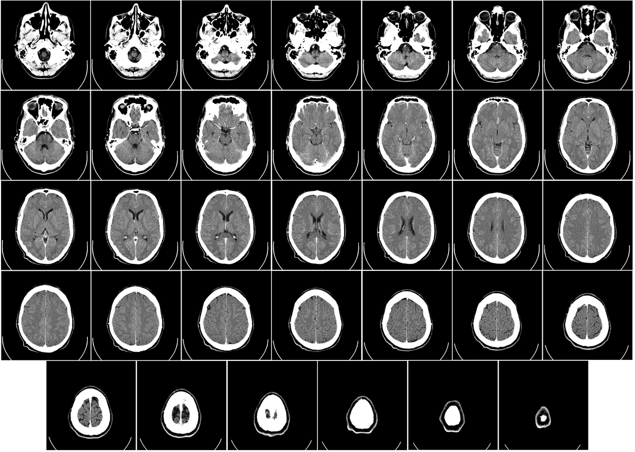
English: Computer tomography of human brain, from base of the skull to top. Taken with intravenous contrast medium.
It was taken Mars 23, 2007 on the uploader, after a 20 minute episode of homonymous hemianopsia with loss of the left visual field, but nothing strange was found. Three episodes of scotoma occurred in the following years, whereof the last one was scintillating (depiction). Otherwise, there were no further neurological symptoms.
Türkçe: Geçirdiği bir kaza neticesinde homonim hemianopsi vakası oluşan bir hastanın beyninin bilgisayarlı tomografisi. Tomografi neticesinde bir anomaliye rastlanmamıştır. |
| 日付 | Uploaded January 17, 2008 |
| 原典 | Radiology, Uppsala University Hospital. Uploaded by Mikael Häggström. |
| 作者 | Department of Radiology, Uppsala University Hospital. Uploaded by Mikael Häggström. |
| 許可 (ファイルの再利用) |
Compound images
[編集]-

-
 Inverted


Scrollable stack
[編集]For larger version, see Category:Computed tomography images of Mikael Häggström's brain. To move through the images, hover over the image and use scroll wheel, drag the mouse, or click the < or the > above each stack. This functionality should activate when the page is fully loaded, which may take some time.

.png)
.png)
.png)
.png)
.png)
.png)
.png)
.png)
.png)
.png)
.png)
.png)
.png)
.png)
.png)
.png)
.png)
.png)
.png)
.png)
.png)
.png)
.png)
.png)
.png)
.png)
.png)
.png)
.png)
.png)
.png)
.png)
.png)
.png)
Case with multiplanar reconstruction
[編集]-
 Brain, case 1: Multiplanar, but no intravenous contrast.
Individual images
[編集]
Licencing
[編集]| このファイルはクリエイティブ・コモンズ CC0 1.0 全世界 パブリック・ドメイン提供のもとで利用可能にされています。 | |
| ある作品に本コモンズ証を関連づけた者は、その作品について世界全地域において著作権法上認められる、その者が持つすべての権利(その作品に関する権利や隣接する権利を含む。)を、法令上認められる最大限の範囲で放棄して、パブリック・ドメインに提供しています。
この作品は、たとえ営利目的であっても、許可を得ずに複製、改変・翻案、配布、上演・演奏することが出来ます。 |
DICOM format
[編集]ファイルの履歴
過去の版のファイルを表示するには、その版の日時をクリックしてください。
| 日付と時刻 | サムネイル | 寸法 | 利用者 | コメント | |
|---|---|---|---|---|---|
| 現在の版 | 2017年12月24日 (日) 01:11 | | 3,639 × 2,595 (3.9メガバイト) | Shashi. (トーク | 投稿記録) | Reverted to version as of 12:49, 1 February 2008 (UTC) |
| 2008年5月8日 (木) 10:59 |  | 3,639 × 2,595 (3.17メガバイト) | CountingPine (トーク | 投稿記録) | Optimise using PNGOUT | |
| 2008年2月1日 (金) 12:49 |  | 3,639 × 2,595 (3.9メガバイト) | Mikael Häggström (トーク | 投稿記録) | {{34 computer tomography images}} {{Individual images of CT of Mikael Häggström's brain}} | |
| 2008年1月31日 (木) 11:56 |  | 3,639 × 2,595 (4.03メガバイト) | Mikael Häggström (トーク | 投稿記録) | {{34 computer tomography images}} {{Individual images of CT of Mikael Häggström's brain}} |
このファイルは上書きできません。
ファイルの使用状況
以下の 41 ページがこのファイルを使用しています:
- User:Dronebogus/Favorites
- User:Mikael Häggström
- File:Anatomy image for main menu.png
- File:CT of brain of Mikael Häggström large.png (リダイレクト)
- File:Computed tomography of human brain (1).png
- File:Computed tomography of human brain (10).png
- File:Computed tomography of human brain (11).png
- File:Computed tomography of human brain (12).png
- File:Computed tomography of human brain (13).png
- File:Computed tomography of human brain (14).png
- File:Computed tomography of human brain (15).png
- File:Computed tomography of human brain (16).png
- File:Computed tomography of human brain (17).png
- File:Computed tomography of human brain (18).png
- File:Computed tomography of human brain (19).png
- File:Computed tomography of human brain (2).png
- File:Computed tomography of human brain (20).png
- File:Computed tomography of human brain (21).png
- File:Computed tomography of human brain (22).png
- File:Computed tomography of human brain (23).png
- File:Computed tomography of human brain (24).png
- File:Computed tomography of human brain (25).png
- File:Computed tomography of human brain (26).png
- File:Computed tomography of human brain (27).png
- File:Computed tomography of human brain (28).png
- File:Computed tomography of human brain (29).png
- File:Computed tomography of human brain (3).png
- File:Computed tomography of human brain (30).png
- File:Computed tomography of human brain (31).png
- File:Computed tomography of human brain (32).png
- File:Computed tomography of human brain (33).png
- File:Computed tomography of human brain (34).png
- File:Computed tomography of human brain (4).png
- File:Computed tomography of human brain (5).png
- File:Computed tomography of human brain (6).png
- File:Computed tomography of human brain (7).png
- File:Computed tomography of human brain (8).png
- File:Computed tomography of human brain (9).png
- File:Computed tomography of human brain - large, inverted.png
- File:Computed tomography of human brain - large.png
- Template:34 computer tomography images
グローバルなファイル使用状況
以下に挙げる他のウィキがこの画像を使っています:
- bn.wikipedia.org での使用状況
- bo.wikipedia.org での使用状況
- ca.wikipedia.org での使用状況
- en.wikipedia.org での使用状況
- CT scan
- Portal:Medicine
- Portal:Medicine/Selected picture
- Portal:Medicine/Selected picture archive
- Wikipedia:WikiProject Neuroscience
- Wikipedia:Featured pictures/Sciences/Biology
- User:Mikael Häggström
- User talk:Mikael Häggström/Archive 1
- Wikipedia:Featured pictures thumbs/10
- Wikipedia:Featured picture candidates/CT of brain of Mikael Häggström.png
- Wikipedia:Featured picture candidates/February-2008
- Wikipedia:Wikipedia Signpost/2008-02-11/Features and admins
- Portal:Medicine/Selected picture/9, 2008
- Portal:Medicine/Selected picture/9
- Wikipedia:Picture of the day/July 2008
- Template:POTD/2008-07-11
- Wikipedia:Wikipedia Signpost/2008-02-11/SPV
- User:Mikael Häggström/Gallery
- Wikipedia:WikiProject Medicine/Recognized content
- Computed tomography of the head
- Wikipedia:Wikipedia Signpost/2013-10-02/Op-ed
- Wikipedia:Wikipedia Signpost/Single/2013-10-02
- User:Wouterstomp/test
- User:Fitness queen04/sandbox
- Wikipedia:WikiProject Anatomy/Resources
- Wikipedia:WikiProject Anatomy/Recognized content
- Wikipedia talk:WikiProject Anatomy/Archive 9
- Reconstruction from projections
- User:VGrigas (WMF)/Quality Media
- User:Flyer22 Frozen/Human brain
- Portal:Medicine/Recognized content
- User talk:Rhododendrites/Reconsidering FPC on the English Wikipedia
- User:KSo007
- en.wikiversity.org での使用状況
- es.wikipedia.org での使用状況
- fi.wikipedia.org での使用状況
- he.wikipedia.org での使用状況
- hy.wikipedia.org での使用状況
- hyw.wikipedia.org での使用状況
- id.wikipedia.org での使用状況
- is.wikipedia.org での使用状況
このファイルのグローバル使用状況を表示する。