File:Development of the notochord in a stage 8 (17–19 days) human embryo.png
Jump to navigation
Jump to search

Size of this preview: 800 × 425 pixels. Other resolutions: 320 × 170 pixels | 640 × 340 pixels | 1,300 × 690 pixels.
Original file (1,300 × 690 pixels, file size: 689 KB, MIME type: image/png)
Captions
Captions
Add a one-line explanation of what this file represents
Summary
[edit]_human_embryo.png&action=edit§ion=1)
| Description |
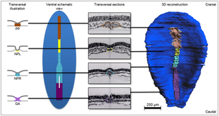
Fig 2. Development of the notochord in a stage 8 (17–19 days) human embryo. |
| Date | |
| Source | https://doi.org/10.1371/journal.pone.0205752 (2018) The development of the human notochord. PLoS ONE 13(10): e0205752. |
| Author | de Bree K, de Bakker BS, Oostra R-J |

|
This file, which was originally posted to an external website, has not yet been reviewed by an administrator or reviewer to confirm that the above license is valid. See Category:License review needed for further instructions.
|
Copyright: © 2018 de Bree et al. This is an open access article distributed under the terms of the Creative Commons Attribution License, which permits unrestricted use, distribution, and reproduction in any medium, provided the original author and source are credited.
Licensing
[edit]_human_embryo.png&action=edit§ion=2)
This file is licensed under the Creative Commons Attribution 4.0 International license.
- You are free:
- to share – to copy, distribute and transmit the work
- to remix – to adapt the work
- Under the following conditions:
- attribution – You must give appropriate credit, provide a link to the license, and indicate if changes were made. You may do so in any reasonable manner, but not in any way that suggests the licensor endorses you or your use.
File history
Click on a date/time to view the file as it appeared at that time.
| Date/Time | Thumbnail | Dimensions | User | Comment | |
|---|---|---|---|---|---|
| current | 21:25, 8 May 2024 | | 1,300 × 690 (689 KB) | Rasbak (talk | contribs) | {{Information |description=Fig 2. Development of the notochord in a stage 8 (17–19 days) human embryo.<br> From left to right transversal illustrations, a schematic ventral view, transversal sections from specimen No. 7545 and a ventral view of a 3D reconstruction of specimen No. 5960. The embryo is viewed from ventral with the endoderm removed. Black lines indicate the level of the transversal sections (epiblast/ectoderm is superior in each transversal section). Subsequent to the gastrulatio... |
You cannot overwrite this file.
File usage on Commons
There are no pages that use this file.
File usage on other wikis
The following other wikis use this file:
- Usage on nl.wikipedia.org
_human_embryo.png&oldid=883330056)