Category:Human embryos
Jump to navigation
Jump to search
first multicellular stage in human development  | |||||
| Upload media | |||||
| Subclass of |
| ||||
|---|---|---|---|---|---|
| Has use |
| ||||
| Followed by | |||||
| Different from | |||||
| Said to be the same as | unborn child | ||||
| |||||
English: In humans, embryo stage start after fertilization and continues untill end of 8th weeks. After this stage, see Category:Foetuses.
Subcategories
This category has the following 6 subcategories, out of 6 total.
A
- Anatomy of human embryos (44 F)
H
- HEK293 cells (189 F)
I
- Illustrations of foetuses (115 F)
T
U
- Ultrasound images of gestational sac (135 F)
Media in category "Human embryos"
The following 188 files are in this category, out of 188 total.
-
 (zh)Human Embryo Day9.png 323 × 330; 60 KB
-
 14 day foetus, 1778 Wellcome L0004602.jpg 1,192 × 1,668; 897 KB
-
 2904 Preembryonic Development-02.jpg 1,957 × 1,792; 1.12 MB
-
 2905 Implantation.jpg 1,950 × 2,440; 1.04 MB
-
 2907 Embroyonic Disc, Amniotic Cavity, Yolk Sac-02-NLtxt.jpg 1,503 × 1,055; 689 KB
-
 2907 Embroyonic Disc, Amniotic Cavity, Yolk Sac-02.jpg 1,529 × 1,055; 551 KB
-
 2908 Germ Layers-02-nltxt.jpg 1,854 × 1,658; 1.45 MB
-
 2908 Germ Layers-02.jpg 1,781 × 1,590; 1.06 MB
-
 2909 Embryo Week 3-02 NLtxt.jpg 1,446 × 1,276; 855 KB
-
 2909 Embryo Week 3-02.jpg 1,447 × 1,252; 634 KB
-
 2913 Embryonic Folding.jpg 1,894 × 1,735; 831 KB
-
 6 weeks pregnant.jpg 120 × 120; 2 KB
-
 6 weeks pregnant.png 120 × 120; 9 KB
-
 819 Embryo at Seven Weeks.jpg 702 × 750; 353 KB
-
 9-Week Human Embryo from Ectopic Pregnancy.jpg 2,054 × 3,081; 2.49 MB
-
 A human embryo of 2 mm. in median sagittal section.jpg 838 × 1,006; 310 KB
-
 Abortus.PNG 199 × 151; 10 KB
-
 Alimentary Canal during the 5th week.jpg 819 × 646; 218 KB
-
 Alimentary Canal in a human embryo of the 3rd week.jpg 967 × 624; 250 KB
-
 Amniotic sac (01).jpg 4,000 × 3,000; 2.6 MB
-
 Approximately 8=10 week EGA from Conception Embryo (21281733395).jpg 1,998 × 1,282; 1.04 MB
-
 Basel 2012-10-05 Batch 2 (6).JPG 447 × 586; 86 KB
-
 Combined.png 206 × 127; 14 KB
-
 Diagram showing human embryo grades for in vitro fertilisation (IVF).jpg 1,699 × 1,139; 212 KB
-

-
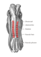
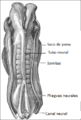 Dorso de embrión humano.png 305 × 452; 48 KB
-
 Early human embryos (27872285595).jpg 1,300 × 1,030; 204 KB
-
 Ecker-Vorlage.jpg 2,462 × 3,320; 3.07 MB
-
 Ectopic pregnancy (01).jpg 3,600 × 2,400; 5.88 MB
-
 Ectopic pregnancy with human embryo.jpg 2,988 × 5,312; 1,020 KB
-

-
 Embryo 3 to 4 weeks in gestational sac (01).jpg 3,568 × 2,379; 3.09 MB
-
 Embryo 3 to 4 weeks in gestational sac.jpg 3,740 × 2,493; 2.6 MB
-
 Embryo 7 weeks after conception.jpg 1,798 × 2,144; 577 KB
-
 Embryo at 4 to 5 weeks fallopian tube (01).jpg 2,000 × 1,334; 1.35 MB
-
 Embryo at 4 to 5 weeks fallopian tube.jpg 1,800 × 2,250; 2.46 MB
-
 Embryo at approximately 8-10 weeks EGA from conception. (21307040921).jpg 1,440 × 1,800; 1,007 KB
-
 Embryo at around 6 weeks EGA from conception (42881289375).jpg 5,447 × 3,632; 10.43 MB
-
 Embryo disc human.jpg 3,326 × 2,096; 449 KB
-
 Embryo in gestational sac.jpg 4,256 × 2,832; 3.87 MB
-
 Embryo week 9-10.jpg 2,103 × 3,155; 2.93 MB
-
 Embryo, 8 cells, transparent image.png 340 × 330; 87 KB
-
 Embryo, 8 cells.jpg 640 × 480; 134 KB
-
 End of week 4 Embryo with somites nltxt.jpg 925 × 674; 203 KB
-
 End of week 4 Embryo with somites.jpg 960 × 720; 121 KB
-
 F.M. van Helmont, Development of human embryo. Wellcome L0011234.jpg 1,114 × 1,726; 609 KB
-
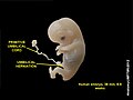
 Formation of the Umbilical Region. human embryo, 1.7 mm. long.jpg 1,065 × 857; 974 KB
-
 Formation of the Umbilicus and Allantois. human embryo, 0.7 mm. long..jpg 1,152 × 803; 970 KB
-
 Four diagrams showing hypothetical stages of early human embryos.jpg 1,631 × 1,434; 943 KB
-
 Germ layers-ar.jpg 1,117 × 264; 151 KB
-
 Germ layers.jpg 1,117 × 264; 229 KB
-
 Gestational sac.svg 603 × 704; 20 KB
-
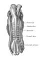
 Gray20.png 304 × 450; 27 KB
-
 Gray20de somite highlight.png 304 × 450; 104 KB
-
 Gray20de.png 304 × 450; 40 KB
-
 Gray20nl somite highlight.png 774 × 1,073; 324 KB
-
 Gray20nl.png 325 × 450; 65 KB
-
 Gray22.png 300 × 303; 23 KB
-
 Gray31.png 400 × 409; 44 KB
-
 Gray32.png 500 × 417; 53 KB
-
 Gray40.png 727 × 838; 383 KB
-
 Gray41.png 300 × 366; 25 KB
-
 Gray44.png 500 × 314; 21 KB
-
 Gray45.png 300 × 379; 24 KB
-
 Gray47.png 222 × 379; 24 KB
-
 Gray59.png 350 × 389; 22 KB
-
 Gray60.png 325 × 309; 21 KB
-
 Gray61.png 382 × 343; 29 KB
-
 Gray62.png 320 × 400; 23 KB
-
 Gray63.png 261 × 450; 36 KB
-
 Gray63.svg 242 × 433; 3 KB
-
 Gray978.png 400 × 497; 42 KB
-
 Hand-book of physiology (1892) (14785304043).jpg 1,792 × 888; 162 KB
-
 Human Embryo (7th week of pregnancy) (304334264).jpg 1,874 × 2,000; 1.36 MB
-
 Human Embryo - (cropped).JPG 1,945 × 1,573; 1.1 MB
-
 Human embryo - 6 weeks.jpg 120 × 120; 5 KB
-
 Human Embryo - Approximately 8 weeks estimated gestational age (1.1).jpg 3,456 × 2,592; 4.52 MB
-
 Human Embryo - Approximately 8 weeks estimated gestational age.jpg 3,872 × 2,592; 4.73 MB
-
 Human embryo 2-ar.JPG 960 × 720; 63 KB
-
 Human embryo 2.JPG 960 × 720; 36 KB
-
 Human embryo 3.JPG 960 × 720; 43 KB
-
 Human Embryo 7 mm. long, Showing the Umbilical Region.jpg 702 × 888; 495 KB
-
 Human embryo 8 weeks 4.JPG 960 × 720; 49 KB
-
 Human embryo 8 weeks 5.JPG 960 × 720; 37 KB
-
 Human embryo 8 weeks 6.JPG 960 × 720; 42 KB
-
 Human embryo 8 weeks 7.JPG 960 × 720; 46 KB
-
 Human embryo 8 weeks 8.JPG 960 × 720; 51 KB
-
 Human embryo 8 weeks 9.JPG 960 × 720; 45 KB
-
 Human embryo and placenta.jpg 756 × 677; 197 KB
-
 Human Embryo Day 9.jpg 780 × 736; 249 KB
-
 Human Embryo Day9.png 323 × 330; 54 KB
-
 Human embryo Section of embryonic rudiment in Peters' ovum (first week).jpg 1,141 × 857; 540 KB
-

-
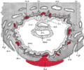
 Human embryo Transverse Section.jpg 932 × 792; 477 KB
-
 Human Embryo.JPG 3,648 × 2,432; 1.52 MB
-
 Human embryo.jpg 960 × 720; 26 KB
-
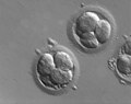
 Human embryos (four-cell stage) (27771482412).jpg 650 × 496; 58 KB
-

-
 Human fetus 10 weeks - therapeutic abortion.jpg 2,592 × 1,944; 1.49 MB
-
 Human foetuses - 5.jpg 2,870 × 4,507; 1.43 MB
-
 Human- Embryo, about 3.5 mm. long.jpg 803 × 791; 701 KB
-
 Human- Embryo, about 5 mm. long.jpg 825 × 846; 809 KB
-
 HumanEmbryogenesis fr j5.svg 512 × 341; 12 KB
-
 HumanEmbryogenesis fr j9.svg 512 × 361; 44 KB
-

-
 Kirkes' handbook of physiology (1907) (14769687492).jpg 1,091 × 918; 435 KB
-
 Lifesize8weekfetus.JPG 400 × 300; 15 KB
-
 Ligne primitive.svg 512 × 406; 55 KB
-
 London NHM 1100625.jpg 2,304 × 3,072; 2.22 MB
-
 Menschlicher-Embryo-8-Wochen Haeckel-1868.jpg 201 × 337; 42 KB
-
 Modell des Kopfes eines menschlichen Fetus am Anfang des 3. Entwicklungsmonats.jpg 1,000 × 1,450; 219 KB
-
 Modell eines implantierten menschlichen Eies am Anfang der 3. Entwicklungswoche.jpg 1,000 × 1,089; 365 KB
-
 Modell eines implantierten menschlichen Eies in der 2. Entwicklungswoche.jpg 1,000 × 1,640; 349 KB
-

-
-

-
-

-

-

-

-

-

-

-

-

-

-
-

-

-
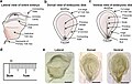
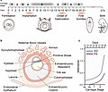
 Morphological differences between human and mouse gastrulation.jpg 2,994 × 3,411; 397 KB
-
 MRM of human embryo at 400MHz.jpg 1,370 × 1,204; 233 KB
-
 MRM of human embryo.jpg 731 × 615; 88 KB
-
 Natural History Museum 306 (8043318253).jpg 3,216 × 4,288; 3.93 MB
-
 Neurula human.png 404 × 529; 112 KB
-
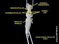
 Pelvix and Lower Spine of Human Embryo (3460140262).jpg 1,469 × 1,632; 1.19 MB
-

-
 Primitive Trilaminar Human Embryo in Tubal Pregnancy (40X) (3944578509).jpg 1,712 × 1,206; 875 KB
-
 PSM V84 D540 Facts and factors of development fig16.jpg 566 × 767; 90 KB
-
 Ruptured cornual ectopic pregnancy.jpg 2,821 × 2,087; 1.08 MB
-
 Ruptured ectopic pregnancy.jpg 3,648 × 2,432; 1.52 MB
-
 Slide1MINI.JPG 960 × 720; 35 KB
-
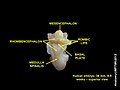
 Spine of Human Embryo (3459324043).jpg 1,426 × 1,564; 935 KB
-
 Staging Human Gastrulation.jpg 3,493 × 3,010; 538 KB
-
 The American journal of anatomy (1901) (14745584956).jpg 1,992 × 2,996; 1.44 MB
-
 The American journal of anatomy (1901) (14766240374).jpg 2,300 × 3,020; 1.18 MB
-
 The American journal of anatomy (1901) (14768266842).jpg 2,526 × 3,588; 883 KB
-
 The American journal of anatomy (1901) (14768274772).jpg 1,968 × 2,996; 1.36 MB
-
 The American journal of anatomy (1901) (14768591405).jpg 2,288 × 3,056; 1.04 MB
-

-

-
 The science and art of midwifery (1891) (14579438820).jpg 2,184 × 3,372; 1.11 MB
-
 The science and art of midwifery (1897) (14576692588).jpg 2,160 × 3,336; 861 KB
-
 Tubal Pregnancy with embryo.jpg 1,874 × 2,000; 1.48 MB
-
 Tubal Pregnancy with Yolk Sac Inside Chorionic Cavity (14961233838).jpg 2,048 × 1,536; 720 KB
-
 Tubal pregnancy, gross pathology 01ee049 lores-2.jpg 637 × 473; 181 KB
-
 Tubal pregnancy, gross pathology 01ee049 lores.jpg 699 × 478; 77 KB
-
 Umbilical Abnormal Structures (01).jpg 761 × 546; 409 KB
-
 Umbilical Abnormal Structures (02).jpg 778 × 562; 393 KB
-
 Umbilical Abnormal Structures.jpg 925 × 691; 538 KB
-
 Umbilical Cord of a Human Embryo 12.5 mm. in length.jpg 1,001 × 1,445; 1.15 MB
-
 Umbilical Cord of a Human Embryo 18 mm. in length.jpg 601 × 779; 537 KB
-
 Umbilical Region in a Human Embryo 10 cm. in length.jpg 971 × 826; 652 KB
-
 Umbilical Region in a Human Embryo 12 cm. in length. (X 3).jpg 652 × 807; 406 KB
-
 Umbilical Region in a Human Embryo 12 cm. in Length.jpg 707 × 707; 585 KB
-
 Umbilical Region in a Human Embryo 12 cm. long.jpg 1,040 × 866; 823 KB
-
 Umbilical Region in a Human Embryo 15 cm. long. (X 4).jpg 857 × 757; 421 KB
-
 Umbilical Region in a Human Embryo 23 mm. in length.jpg 960 × 833; 729 KB
-
 Umbilical Region in a Human Embryo 4.5 cm. in length.jpg 905 × 1,383; 854 KB
-
 Umbilical Region in a Human Embryo 7.5 cm. long.jpg 777 × 691; 432 KB
-
 Umbilical Region in a Human Embryo 9 cm. in length.jpg 873 × 856; 560 KB
-
 Umbilical Region in an Embryo 7 mm. in length.jpg 882 × 713; 492 KB
-
 Umbilical Region of a Fetus at Term. Abnormal.jpg 613 × 1,116; 498 KB
-
 Umbilical Region of a Human Embryo 10 mm. in length.jpg 1,167 × 957; 1.09 MB
-
 Umbilical Region of a Human Embryo 3 cm. long.jpg 804 × 769; 769 KB
-

-
 Umbilical Region of a Human Embryo 5.2 cm. in Length.jpg 864 × 945; 763 KB
-
 Umbilical Region of a Human Embryo 6.5 cm. in length.jpg 684 × 898; 594 KB
-
 Umbilicus in a Human Embryo 12 cm. in length. (X 8).jpg 629 × 801; 538 KB
-
 Views of a Foetus in the Womb.jpg 996 × 1,063; 657 KB
-
 Yolk-sac with the embryo 5.5 cm.jpg 718 × 575; 362 KB
-
 Zn 46 hrs 2-18-09.jpg 220 × 221; 32 KB
-
 Zn46 DAPI 2-18-09.jpg 206 × 206; 26 KB
-
 Бластоциста человека 5-е сутки развития.jpg 548 × 477; 445 KB
-
 Эмбрион человека 2-е сутки развития.jpg 445 × 428; 336 KB
-
 Эмбрион человека 3-и сутки развития (8сell).png 501 × 443; 456 KB
-
 Эмбрион человека на 4-е сутки развития.jpg 528 × 419; 430 KB
-
 Эмбрион человека первые сутки развития.png 408 × 407; 355 KB
Human_Embryo_Day9.png)


.jpg)
.jpg)
.JPG)

.jpg)
_025_Hinteres_K%C3%B6rperende_einer_Leibesfrucht.png)

.jpg)

.jpg)


.jpg)


.jpg)

.jpg)
.jpg)


_(14785304043).jpg)
_(304334264).jpg)
.JPG)

.jpg)


.jpg)


_(27771482412).jpg)


_(14598556478).jpg)
_(14769687492).jpg)


_am_Anfang_des_2._Entwicklungsmonats_(2).jpg)
_am_Anfang_des_2._Entwicklungsmonats.jpg)
_(2).jpg)
_(5).jpg)
_(2).jpg)
_(5).jpg)
_(6).jpg)
_(2).jpg)
_(3).jpg)
_(2).jpg)
_(2).jpg)
_(5).jpg)
_(6).jpg)
_(4).jpg)
_(5).jpg)
_(7).jpg)
_(3).jpg)


.jpg)

.jpg)
_(14577936487).jpg)
_(3944578509).jpg)


.jpg)

_(14745584956).jpg)
_(14766240374).jpg)
_(14768266842).jpg)
_(14768274772).jpg)
_(14768591405).jpg)
_(14781412895).jpg)
_(14776060862).jpg)
_(14579438820).jpg)
_(14576692588).jpg)

.jpg)


.jpg)
.jpg)


.jpg)


.jpg)


.jpg)


.png)

