File:Diagrams and images of human embryos at the gastrula stage.png
Jump to navigation
Jump to search

Size of this preview: 587 × 599 pixels. Other resolutions: 235 × 240 pixels | 470 × 480 pixels | 752 × 768 pixels | 1,003 × 1,024 pixels | 2,006 × 2,048 pixels | 3,128 × 3,193 pixels.
Original file (3,128 × 3,193 pixels, file size: 804 KB, MIME type: image/png)
Captions
Captions
Add a one-line explanation of what this file represents
Summary[edit]
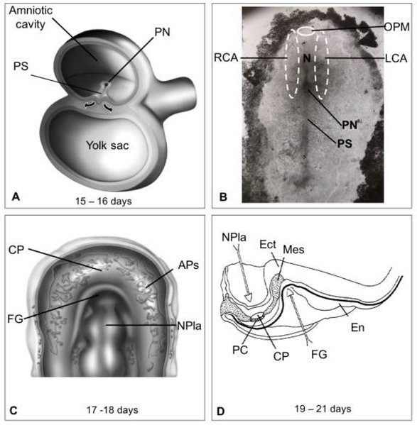
| Description | Figure 2. Diagrams and images of embryos at the gastrula stage. (A) Development of the trilaminar embryonic disk of chicken due to the migration (arrows) of epiblast cells through the primitive streak (PS). (B) A 16 ± 1-day embryo exhibiting the right (RCA) and left (LCA) cardiogenic areas spanning one-third of the primitive streak, the primitive node (PN), and the notochord (N) to the oropharyngeal membrane (OPM). (C) An 18 ± 1-day embryo showing the angiogenic plexuses (APs) arranged in the cardiac crescent, facing the neural plate (NPla). (D) Sagittal section of a 19–21-day human embryo with the first pair of somites showing the beginning of the development of the foregut (FG) and the neural plate. The somatopleure (SP), pericardial cavity (PC), and cardiogenic plate (CP) on the endoderm (En) of the yolk sac are also visible |
| Date | |
| Source | https://doi.org/10.3390/life13010165 Human Heart Morphogenesis: A New Vision Based on In Vivo Labeling and Cell Tracking. Life 2023, 13, 165. |
| Author | Villavicencio-Guzmán, L.; Sánchez-Gómez, C.; Jaime-Cruz, R.; Ramírez-Fuentes, T.C.; Patiño-Morales, C.C.; Salazar-García, M. |

|
This file, which was originally posted to an external website, has not yet been reviewed by an administrator or reviewer to confirm that the above license is valid. See Category:License review needed for further instructions.
|
© 2023 by the authors. Licensee MDPI, Basel, Switzerland. This article is an open access article distributed under the terms and conditions of the Creative Commons Attribution (CC BY) license (https://creativecommons.org/licenses/by/4.0/).
Licensing[edit]
This file is licensed under the Creative Commons Attribution 4.0 International license.
- You are free:
- to share – to copy, distribute and transmit the work
- to remix – to adapt the work
- Under the following conditions:
- attribution – You must give appropriate credit, provide a link to the license, and indicate if changes were made. You may do so in any reasonable manner, but not in any way that suggests the licensor endorses you or your use.
File history
Click on a date/time to view the file as it appeared at that time.
| Date/Time | Thumbnail | Dimensions | User | Comment | |
|---|---|---|---|---|---|
| current | 00:22, 5 May 2024 | | 3,128 × 3,193 (804 KB) | Rasbak (talk | contribs) | {{Information |description= Figure 2. Diagrams and images of embryos at the gastrula stage. (A) Development of the trilaminar embryonic disk of chicken due to the migration (arrows) of epiblast cells through the primitive streak (PS). (B) A 16 ± 1-day embryo exhibiting the right (RCA) and left (LCA) cardiogenic areas spanning one-third of the primitive streak, the primitive node (PN), and the notochord (N) to the oropharyngeal membrane (OPM). (C) An 18 ± 1-day embryo showing the angiogenic pl... |
You cannot overwrite this file.
File usage on Commons
There are no pages that use this file.
File usage on other wikis
The following other wikis use this file:
- Usage on nl.wikipedia.org