File:European foulbrood BHL41863898.jpg

Original file (2,628 × 4,260 pixels, file size: 1.68 MB, MIME type: image/jpeg)
Captions
Captions
Summary
[edit]| Description |
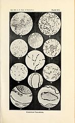
Plate VII Photomicrographs illustrating the more commonly encountered bacteria in European foulbrood. A. — Bacillus pluton: A smear from the stomach of a larva sick with European foulbrood. Note the paired forms and short chains. These forms are numerous in a recent infection, suggesting the organism in the process of multiplication. The lancet-shaped form is by far the predominant one in all later stages of the disease. X 1000. B. — Bacillus pluton: A smear from a larva quite recently infected. The multiplying paired forms are at this stage present almost exclusively. X 1000. C. — Bacterium eurydice: Stained preparation from a pure culture on the surface of agar. X 1000. D. — Bacillus alvei: Stained preparation showing spores and spore formation. X 800. E. — Streptococcus apis: Stained preparation from a pure culture. X 800. F. — Bacillus alvei: The peculiar arrangement of the spores as sometimes seen. From a pure culture, the smear having been made by suspending the culture on the slide in normal salt solution. X 1000. G. — Bacillus orpheus: Stained preparation made from a pure culture only a few hours old. Grown on the surface of agar. X 1000. H. — Bacillus orpheus: Stained preparation showing spore formation. Note the stained portion along one side and about both ends of the spore. The stage is soon reached in a culture at incubator temperature. At room temperature it remains in this stage for a considerable period. X 800. I. — Longisection of a young larva showing early infection in European foulbrood. The bacterial growth is seen as a narrow black area just within the peritrophic membrane on one side of the food mass. J. — Longisection of larva sick of European foulbrood. showing a later stage of infection than that present in I. The dark area in the food mass shows the bacterial growth. Note that the growth mass does not extend beyond the peritrophic membrane and that it does not extend uniformly along this membrane and throughout the food mass. K. — Transverse section of larva about the time of its death from European foulbrood infection. Note the bacterial mass along the peritrophic membrane and extending from the membrane into the food mass. As seen within the living larva this bacterial mass in the sick larva is practically white, but is more or less yellowish white when present with larval food material. The gelatinous-like envelope outside the peritrophic membrane and inside the stomach epithelium in healthy larvae thins out as the disease advances. |
||
| Date | |||
| Source | https://www.biodiversitylibrary.org/pageimage/41863898 | ||
| Author | White, G. F. | ||
| Page ID | 41863898 | ||
| Item ID | 131054 (Find related Wikimedia Commons images) | ||
| Title ID | 64650 (Find related Wikimedia Commons images) | ||
| BHL Page URL | https://www.biodiversitylibrary.org/page/41863898 | ||
| DOI | 10.5962/bhl.title.64650 | ||
| Page type | Text Illustration | ||
| Credit | The contributing institution believes that this item is not in copyright
|

Licensing
[edit]
This image is in the public domain because it is a mere mechanical scan or photocopy of a public domain original, or – from the available evidence – is so similar to such a scan or photocopy that no copyright protection can be expected to arise. The original itself is in the public domain for the following reason:
This tag is designed for use where there may be a need to assert that any enhancements (eg brightness, contrast, colour-matching, sharpening) are in themselves insufficiently creative to generate a new copyright. It can be used where it is unknown whether any enhancements have been made, as well as when the enhancements are clear but insufficient. For known raw unenhanced scans you can use an appropriate {{PD-old}} tag instead. For usage, see Commons:When to use the PD-scan tag.  | ||||
File history
Click on a date/time to view the file as it appeared at that time.
| Date/Time | Thumbnail | Dimensions | User | Comment | |
|---|---|---|---|---|---|
| current | 14:29, 2 November 2015 | | 2,628 × 4,260 (1.68 MB) | Fæ (talk | contribs) | == {{int:filedesc}} == {{BHL | source = http://www.biodiversitylibrary.org/pageimage/41863898 | description = European foulbrood / | pageid = 41863898 | itemid = 131054 | titleid = 64650 | pagenumbers = | pagetypes = Text Illustration | volume = no.8... |
You cannot overwrite this file.
File usage on Commons
There are no pages that use this file.
File usage on other wikis
The following other wikis use this file:
- Usage on de.wikipedia.org
- Usage on es.wikipedia.org