File:Free-living amebic infections.png

Alkuperäinen tiedosto (1 365 × 1 170 kuvapistettä, 715 KiB, MIME-tyyppi: image/png)
Kuvatekstit
Kuvatekstit

|
Tämä tyyppiä biology oleva kuva pitäisi luoda uudelleen SVG-tiedostoksi vektorigrafiikan avulla. Tällä tiedostotyypillä on monia vahvuuksia, sivulta Commons:Media for cleanup löytyy lisätietoja. Jos tästä kuvasta on jo olemassa SVG-versio, ole ystävällinen ja tallenna se tänne. SVG-tiedoston tallentamisen jälkeen vaihda tämä malline mallineeseen {{vector version available|uusi kuvan nimi.svg}}.
|
Yhteenveto[muokkaa]
| KuvausFree-living amebic infections.png |
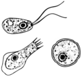
English: This is an illustration of the life cycle of the parasitic agents responsible for causing “free-living” amebic infections.
For a complete description of the life cycle of these parasites, select the link below the image or paste the following address in your address bar: http://www.dpd.cdc.gov/dpdx/HTML/FreeLivingAmebic.htm Free-living amebae belonging to the genera Acanthamoeba, Balamuthia, and Naegleria are important causes of disease in humans and animals. Naegleria fowleri produces an acute, and usually lethal, central nervous system (CNS) disease called primary amebic meningoencephalitis (PAM). N. fowleri has three stages, cysts (1) , trophozoites (2) , and flagellated forms (3) , in its life cycle. The trophozoites replicate by promitosis (nuclear membrane remains intact) (4) . Naegleria fowleri is found in fresh water, soil, thermal discharges of power plants, heated swimming pools, hydrotherapy and medicinal pools, aquariums, and sewage. Trophozoites can turn into temporary flagellated forms which usually revert back to the trophozoite stage. Trophozoites infect humans or animals by entering the olfactory neuroepithelium (5) and reaching the brain. N. fowleri trophozoites are found in cerebrospinal fluid (CSF) and tissue, while flagellated forms are found in CSF. Acanthamoeba spp. and Balamuthia mandrillaris are opportunistic free-living amebae capable of causing granulomatous amebic encephalitis (GAE) in individuals with compromised immune systems. Acanthamoeba spp. have been found in soil; fresh, brackish, and sea water; sewage; swimming pools; contact lens equipment; medicinal pools; dental treatment units; dialysis machines; heating, ventilating, and air conditioning systems; mammalian cell cultures; vegetables; human nostrils and throats; and human and animal brain, skin, and lung tissues. B. mandrillaris however, has not been isolated from the environment but has been isolated from autopsy specimens of infected humans and animals. Unlike N. fowleri, Acanthamoeba and Balamuthia have only two stages, cysts (1) and trophozoites (2) , in their life cycle. No flagellated stage exists as part of the life cycle. The trophozoites replicate by mitosis (nuclear membrane does not remain intact) (3) . The trophozoites are the infective forms and are believed to gain entry into the body through the lower respiratory tract, ulcerated or broken skin and invade the central nervous system by hematogenous dissemination (4). Acanthamoeba spp. and Balamuthia mandrillaris cysts and trophozoites are found in tissue. |
||
| Päiväys | |||
| Lähde |
|
||
| Tekijä |
|
||
| Käyttöoikeus (Tämän tiedoston uudelleenkäyttö) |
English: None - This image is in the public domain and thus free of any copyright restrictions. As a matter of courtesy we request that the content provider be credited and notified in any public or private usage of this image. |
||
| Muut versiot |
|
Lisenssi[muokkaa]
| Public domainPublic domainfalsefalse |
Tämän teoksen on valmistanut Yhdysvaltain tartuntatautien valvonta- ja ehkäisykeskusten (Centers for Disease Control and Prevention, CDC) työntekijä osana kyseisen työntekijän virkatointa. Yhdysvaltain liittovaltion viranomaisten työntekijöiden tekemät teokset eivät saa tekijänoikeuden suojaa Yhdysvaltain tekijänoikeuslain 105 § mukaisesti.
eesti ∙ Deutsch ∙ čeština ∙ español ∙ português ∙ English ∙ français ∙ Nederlands ∙ polski ∙ slovenščina ∙ suomi ∙ македонски ∙ українська ∙ 日本語 ∙ 中文(简体) ∙ 中文(繁體) ∙ العربية ∙ +/− |
Alkuperäinen tallennusloki[muokkaa]
Siirretty projektista en.wikipedia Commonsiin käyttäjän Optigan13 toimesta.
- 2006-05-04 01:09 Keenan Pepper 518×435×8 (31658 bytes) Free-living_amebic_infections.gif, converted to [[PNG]] by [[netpbm]].
Tiedoston historia
Päiväystä napsauttamalla näet, millainen tiedosto oli kyseisellä hetkellä.
| Päiväys | Pienoiskuva | Koko | Käyttäjä | Kommentti | |
|---|---|---|---|---|---|
| nykyinen | 2. helmikuuta 2023 kello 09.24 |  | 1 365 × 1 170 (715 KiB) | Materialscientist (keskustelu | muokkaukset) | https://answersingenesis.org/biology/microbiology/the-genesis-of-brain-eating-amoeba/ |
| 20. heinäkuuta 2008 kello 06.30 |  | 518 × 435 (31 KiB) | Optigan13 (keskustelu | muokkaukset) | {{Information |Description={{en|This is an illustration of the life cycle of the parasitic agents responsible for causing “free-living” amebic infections. For a complete description of the life cycle of these parasites, select the link below the image |
Et voi tallentaa uutta tiedostoa tämän tilalle.
Tiedoston käyttö
Seuraavat 2 sivua käyttävät tätä tiedostoa:
Tiedoston järjestelmänlaajuinen käyttö
Seuraavat muut wikit käyttävät tätä tiedostoa:
- Käyttö kohteessa de.wikibooks.org
- Käyttö kohteessa en.wiktionary.org
- Käyttö kohteessa fi.wikipedia.org
- Käyttö kohteessa fr.wikipedia.org
- Käyttö kohteessa gl.wikipedia.org
- Käyttö kohteessa hr.wikipedia.org
- Käyttö kohteessa is.wikipedia.org
- Käyttö kohteessa it.wikipedia.org
- Käyttö kohteessa pl.wikipedia.org
- Käyttö kohteessa te.wikipedia.org
- Käyttö kohteessa vi.wikipedia.org
- Käyttö kohteessa www.wikidata.org
- Käyttö kohteessa zh.wikipedia.org
Metatieto
Tämä tiedosto sisältää esimerkiksi kuvanlukijan, digikameran tai kuvankäsittelyohjelman lisäämiä lisätietoja. Kaikki tiedot eivät enää välttämättä vastaa todellisuutta, jos kuvaa on muokattu sen alkuperäisen luonnin jälkeen.
| Kuvan resoluutio leveyssuunnassa | 28,35 dpc |
|---|---|
| Kuvan resoluutio korkeussuunnassa | 28,35 dpc |
| Viimeksi muokattu | 2. helmikuuta 2023 kello 09.24 |


