File:Histology of apocrine cells.png
Jump to navigation
Jump to search

Size of this preview: 509 × 600 pixels. Other resolutions: 204 × 240 pixels | 407 × 480 pixels | 652 × 768 pixels | 1,189 × 1,401 pixels.
Original file (1,189 × 1,401 pixels, file size: 1.7 MB, MIME type: image/png)
Captions
Captions
Add a one-line explanation of what this file represents
Summary[edit]
| Description |
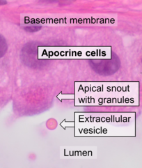
English: Histology of apocrine cells, H&E stain. From a case of apocrine metaplasia of breast glands. |
| Date | |
| Source | Own work |
| Author |
.jpg) - Reusing images - Conflicts of interest: None Consent note: Consent from the patient or patient's relatives is regarded as redundant, because of absence of identifiable features (List of HIPAA identifiers) in the media and case information (See also HIPAA case reports guidance). |
| Other versions |
Source images: |


Licensing[edit]
| This file is made available under the Creative Commons CC0 1.0 Universal Public Domain Dedication. | |
| The person who associated a work with this deed has dedicated the work to the public domain by waiving all of their rights to the work worldwide under copyright law, including all related and neighboring rights, to the extent allowed by law. You can copy, modify, distribute and perform the work, even for commercial purposes, all without asking permission.
|
File history
Click on a date/time to view the file as it appeared at that time.
| Date/Time | Thumbnail | Dimensions | User | Comment | |
|---|---|---|---|---|---|
| current | 17:51, 4 February 2024 | | 1,189 × 1,401 (1.7 MB) | Mikael Häggström (talk | contribs) | Showing basement membrane better |
| 17:45, 4 February 2024 |  | 1,197 × 1,441 (1.74 MB) | Mikael Häggström (talk | contribs) | Better | |
| 17:35, 4 February 2024 |  | 1,221 × 1,434 (1.76 MB) | Mikael Häggström (talk | contribs) | Uploaded own work with UploadWizard |
You cannot overwrite this file.
File usage on Commons
The following page uses this file:
File usage on other wikis
The following other wikis use this file:
- Usage on en.wikipedia.org