File:Histopathology of Barrett's esophagus, annotated.jpg
Jump to navigation
Jump to search
Size of this preview: 682 × 599 pixels. Other resolutions: 273 × 240 pixels | 546 × 480 pixels | 874 × 768 pixels | 1,165 × 1,024 pixels | 2,047 × 1,799 pixels.
Original file (2,047 × 1,799 pixels, file size: 828 KB, MIME type: image/jpeg)
Captions
Captions
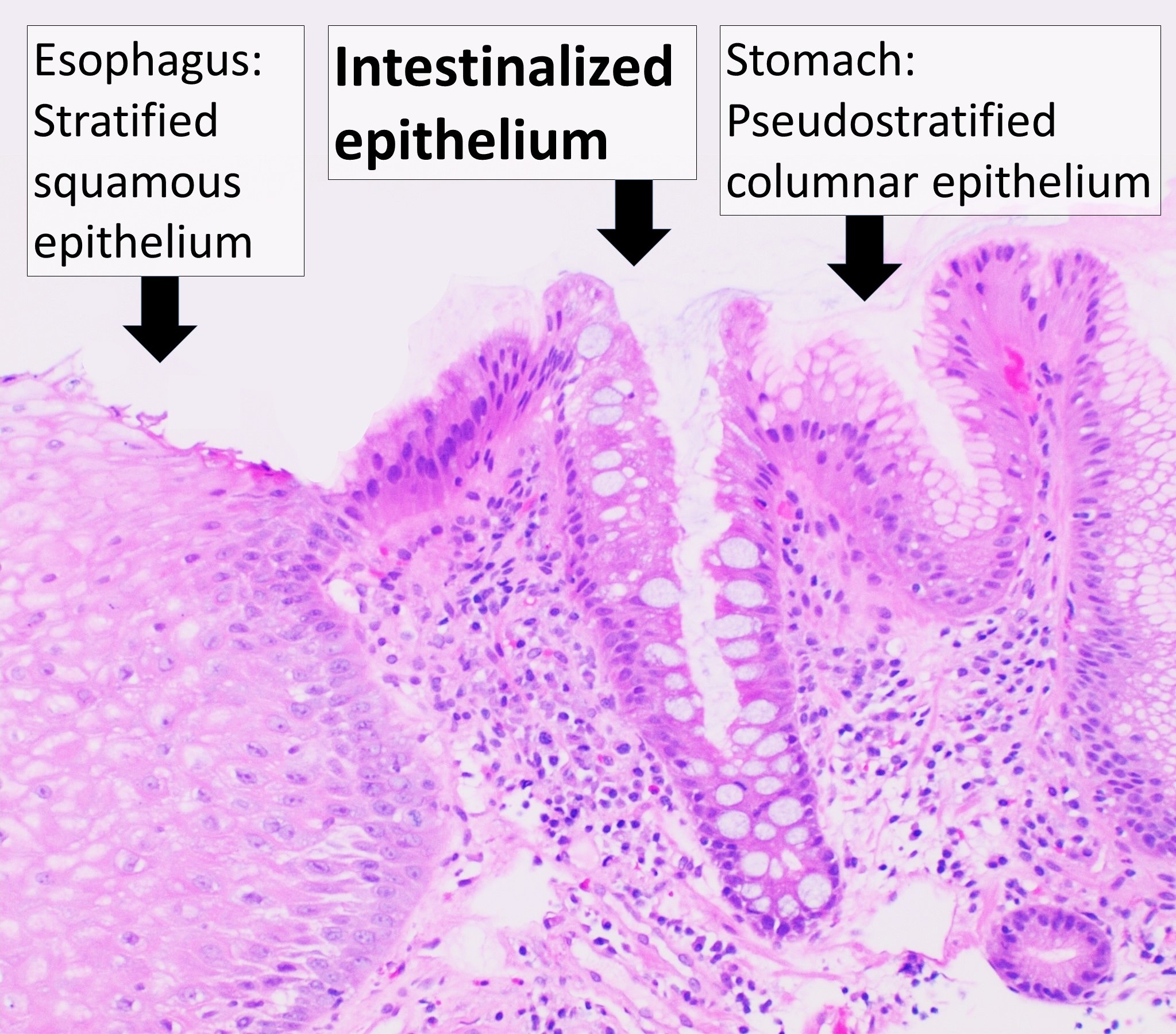
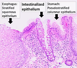
Histopathology of Barrett's esophagus, annotated
Summary
[edit]
| Description |
English: Histopathology of Barrett's esophagus, showing intestinalized epithelium with goblet cells, as opposed to normal stratified squamous epithelium of the esophagus, and pseudostratified columnar epithelium of the fundus of the stomach. The submucosa displays an infiltrate including lymphocytes and plasma cells, constituting an underlying chronic inflammation. The area between the stratified and the intestinalized epithelium displays reactive changes, but there is no secondary dysplasia in this case. H&E stain. |
| Date | |
| Source | Own work |
| Author |
.jpg) - Reusing images - Conflicts of interest: None Consent note: Consent from the patient or patient's relatives is regarded as redundant, because of absence of identifiable features (List of HIPAA identifiers) in the media and case information (See also HIPAA case reports guidance). |
| Other versions |
 |
Licensing
[edit]
| This file is made available under the Creative Commons CC0 1.0 Universal Public Domain Dedication. | |
| The person who associated a work with this deed has dedicated the work to the public domain by waiving all of their rights to the work worldwide under copyright law, including all related and neighboring rights, to the extent allowed by law. You can copy, modify, distribute and perform the work, even for commercial purposes, all without asking permission.
|
File history
Click on a date/time to view the file as it appeared at that time.
| Date/Time | Thumbnail | Dimensions | User | Comment | |
|---|---|---|---|---|---|
| current | 21:26, 3 April 2021 | | 2,047 × 1,799 (828 KB) | Mikael Häggström (talk | contribs) | Spelling error |
| 20:58, 3 April 2021 |  | 2,047 × 1,795 (830 KB) | Mikael Häggström (talk | contribs) | Uploaded a work by {{Mikael Häggström|cat=Micrographs of the gastrointestinal tract|consent=noid}} from {{Own}} with UploadWizard |
You cannot overwrite this file.
File usage on Commons
The following page uses this file:
File usage on other wikis
The following other wikis use this file:
- Usage on en.wikipedia.org