File:Histopathology of goblet cells and foveolar cells in incomplete Barrett's esophagus.jpg
Jump to navigation
Jump to search
Size of this preview: 800 × 598 pixels. Other resolutions: 320 × 239 pixels | 640 × 479 pixels | 1,024 × 766 pixels | 1,280 × 958 pixels | 2,048 × 1,532 pixels.
Original file (2,048 × 1,532 pixels, file size: 665 KB, MIME type: image/jpeg)
Captions
Captions
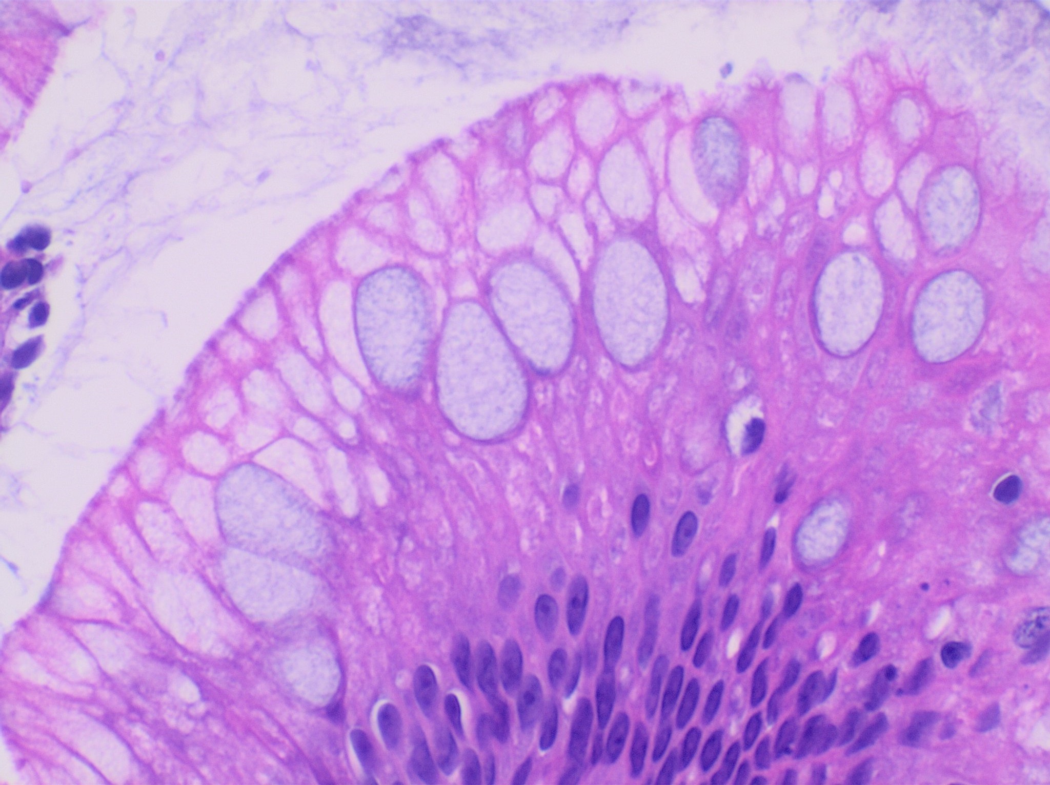
Histopathology of goblet cells in Barrett's esophagus
Summary[edit]
| Description |
English: In incomplete intestinal metaplasia (incomplete Barrett's esophagus), there are both foveolar cells and goblet cells, the latter usually having a slightly bluish color compared to the apical cytoplasm of foveolar cells on H&E stain. |
| Date | |
| Source | Own work |
| Author |
.jpg) - Reusing images - Conflicts of interest: None Consent note: Consent from the patient or patient's relatives is regarded as redundant, because of absence of identifiable features (List of HIPAA identifiers) in the media and case information (See also HIPAA case reports guidance). |
| Other versions |
_and_foveolar_cells_in_incomplete_Barrett%27s_esophagus.jpg) |
Licensing[edit]
| This file is made available under the Creative Commons CC0 1.0 Universal Public Domain Dedication. | |
| The person who associated a work with this deed has dedicated the work to the public domain by waiving all of their rights to the work worldwide under copyright law, including all related and neighboring rights, to the extent allowed by law. You can copy, modify, distribute and perform the work, even for commercial purposes, all without asking permission.
|
File history
Click on a date/time to view the file as it appeared at that time.
| Date/Time | Thumbnail | Dimensions | User | Comment | |
|---|---|---|---|---|---|
| current | 22:59, 30 November 2020 | | 2,048 × 1,532 (665 KB) | Mikael Häggström (talk | contribs) | Uploaded a work by {{Mikael Häggström|cat=Micrographs of the gastrointestinal tract|consent=noid}} from {{Own}} with UploadWizard |
You cannot overwrite this file.
File usage on Commons
The following 2 pages use this file: