File:Histopathology of tubular carcinoma, low magnification.jpg
Jump to navigation
Jump to search
Size of this preview: 800 × 598 pixels. Other resolutions: 320 × 239 pixels | 640 × 479 pixels | 1,024 × 766 pixels | 1,280 × 958 pixels | 2,048 × 1,532 pixels.
Original file (2,048 × 1,532 pixels, file size: 1.12 MB, MIME type: image/jpeg)
Captions
Captions
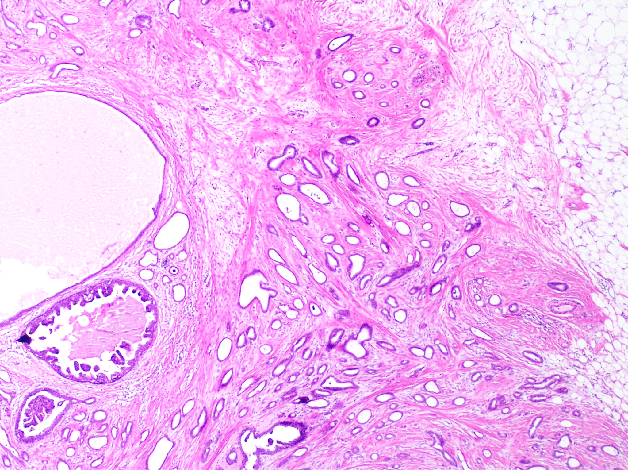
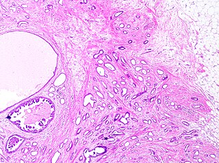
Histopathology of tubular carcinoma, low magnification, H&E stain.
Summary[edit]
| Description |
English: Histopathology of tubular carcinoma, low magnification, H&E stain. It shows the typical features of invasive breast cancer with infiltrative growth pattern, here including invasion into adipose tissue and with an associated fibrous or desmoplastic stromal response. It has the criterion of more than 90% of the tumor composed of small, ovoid or angulated tubules with open lumina. Ductal carcinoma in situ is seen at left as lumina with micropapillary formations (under the larger bland cyst), and was presumably the precursor lesion for the ductal carcinoma.
|
| Date | |
| Source | Own work |
| Author |
.jpg) - Reusing images - Conflicts of interest: None |
| Other versions |
 |
Licensing[edit]
| This file is made available under the Creative Commons CC0 1.0 Universal Public Domain Dedication. | |
| The person who associated a work with this deed has dedicated the work to the public domain by waiving all of their rights to the work worldwide under copyright law, including all related and neighboring rights, to the extent allowed by law. You can copy, modify, distribute and perform the work, even for commercial purposes, all without asking permission.
|
File history
Click on a date/time to view the file as it appeared at that time.
| Date/Time | Thumbnail | Dimensions | User | Comment | |
|---|---|---|---|---|---|
| current | 17:21, 16 October 2023 | | 2,048 × 1,532 (1.12 MB) | Mikael Häggström (talk | contribs) | Uploaded a work by {{Mikael Häggström|cat=Micrographs of the breast}} from {{Own}} with UploadWizard |
You cannot overwrite this file.
File usage on Commons
The following page uses this file:
File usage on other wikis
The following other wikis use this file:
- Usage on en.wikipedia.org