File:Propagation of action potential along myelinated nerve fiber en.svg
Aller à la navigation
Aller à la recherche

Taille de cet aperçu PNG pour ce fichier SVG : 800 × 460 pixels. Autres résolutions : 320 × 184 pixels | 640 × 368 pixels | 1 024 × 589 pixels | 1 280 × 736 pixels | 2 560 × 1 472 pixels | 1 082 × 622 pixels.
Fichier d’origine (Fichier SVG, nominalement de 1 082 × 622 pixels, taille : 120 kio)
Légendes
Légendes
Ajoutez en une ligne la description de ce que représente ce fichier
Description
[modifier]| Description |
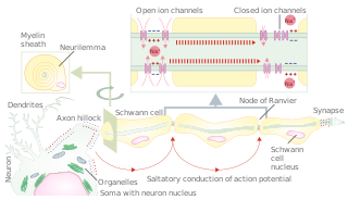
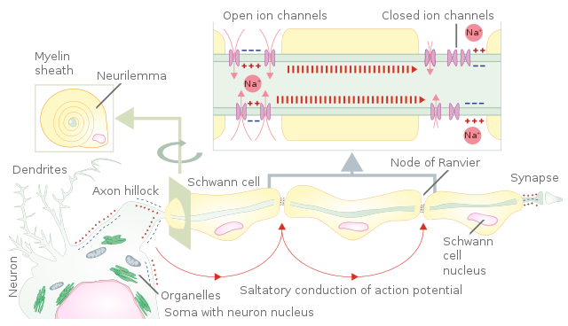
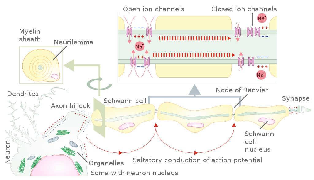
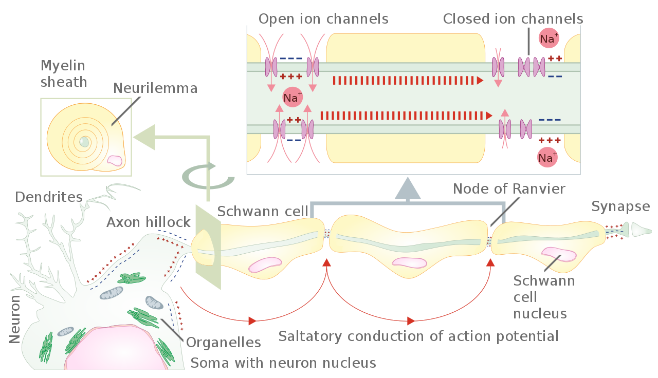
Українська: Переклад англійською File:Propagation of action potential along myelinated nerve fiber.png English: Translation and vector of the File:Propagation of action potential along myelinated nerve fiber.png Image description: Schematic representation of the action potential propagation through myelinated nerve fiber of peripheral nervous system. From axon hillock of neuron body (soma) action potential propagates from one unmyelinated fiber part to the next one. The unmyelinated parts of the nerve fiber are nodes of Ranvier. This way of action potential propagation is called saltatory conduction (red arrows in the diagram) Ion channels open, allow sodium ions to enter the cell leading to membrane depolarization and generation of action potential. Myelination of nerve fibers in the peripheral nervous system is achieved by Schwann cells wrapping around an axon part (cross section). The nucleus and most of the Schwan cell cytoplasm are contained in the outer most layer called neurilemma. Own work loosely based on Raphael Alya R., Talbot William S. (2011). "New Insights into Signaling During Myelination in Zebrafish". Current topics in developmental biology 97: 1–19. DOI:10.1016/B978-0-12-385975-4.00007-3. ISSN 00702153.Min Y., Kristiansen K., Boggs J. M. at al (2009). "Interaction forces and adhesion of supported myelin lipid bilayers modulated by myelin basic protein". Proceedings of the National Academy of Sciences 106 (9): 3154–3159. DOI:10.1073/pnas.0813110106. ISSN 0027-8424. |
| Date | |
| Source | Travail personnel |
| Auteur | Helixitta |
| Autres versions |
|

|
Conditions d’utilisation
[modifier]
Moi, en tant que détenteur des droits d’auteur sur cette œuvre, je la publie sous la licence suivante :
Ce fichier est sous la licence Creative Commons Attribution – Partage dans les Mêmes Conditions 4.0 International.
- Vous êtes libre :
- de partager – de copier, distribuer et transmettre cette œuvre
- d’adapter – de modifier cette œuvre
- Sous les conditions suivantes :
- paternité – Vous devez donner les informations appropriées concernant l'auteur, fournir un lien vers la licence et indiquer si des modifications ont été faites. Vous pouvez faire cela par tout moyen raisonnable, mais en aucune façon suggérant que l’auteur vous soutient ou approuve l’utilisation que vous en faites.
- partage à l’identique – Si vous modifiez, transformez, ou vous basez sur cette œuvre, vous devez distribuer votre contribution sous la même licence ou une licence compatible avec celle de l’original.
| Cette image a été téléversée dans le cadre du Concours de photos scientifiques européen de 2015. |
Historique du fichier
Cliquer sur une date et heure pour voir le fichier tel qu'il était à ce moment-là.
| Date et heure | Vignette | Dimensions | Utilisateur | Commentaire | |
|---|---|---|---|---|---|
| actuel | 18 octobre 2022 à 09:24 | | 1 082 × 622 (120 kio) | Chandres (d | contributions) | File uploaded using svgtranslate tool (https://svgtranslate.toolforge.org/). Added translation for fr. |
| 11 juillet 2016 à 14:19 |  | 1 082 × 622 (116 kio) | Helixitta (d | contributions) | font fix, error fix (?) | |
| 8 juillet 2016 à 21:30 |  | 1 082 × 622 (91 kio) | Helixitta (d | contributions) | transparency issues | |
| 8 juillet 2016 à 21:26 |  | 1 082 × 641 (93 kio) | Helixitta (d | contributions) | fixed text | |
| 8 juillet 2016 à 21:21 |  | 1 082 × 641 (106 kio) | Helixitta (d | contributions) | User created page with UploadWizard |
Vous ne pouvez pas remplacer ce fichier.
Utilisations locales du fichier
Les 14 pages suivantes utilisent ce fichier :
- User:Magog the Ogre/Multilingual legend/2022 October 11-20
- User talk:Helixitta
- Commons:Featured picture candidates/File:Propagation of action potential along myelinated nerve fiber en.svg
- Commons:Featured picture candidates/Log/July 2016
- Commons:Featured pictures/Non-photographic media/Computer-generated
- Commons:Featured pictures/chronological/2016-B
- Commons:Picture of the Year/2016/Candidates
- Commons:Picture of the Year/2016/R1/Gallery/2016-B
- Commons:Picture of the Year/2016/R1/Gallery/ALL
- Commons:Picture of the Year/2016/R1/Gallery/M07
- Commons:Picture of the Year/2016/R1/Gallery/Maps
- Commons:Picture of the Year/2016/R1/v/Propagation of action potential along myelinated nerve fiber en.svg
- File:Propagation of action potential along myelinated nerve fiber.png
- File:Saltatorische Erregungsleitung.svg
Utilisations du fichier sur d’autres wikis
Les autres wikis suivants utilisent ce fichier :
- Utilisation sur ar.wikipedia.org
- Utilisation sur ca.wikipedia.org
- Utilisation sur cs.wikipedia.org
- Utilisation sur en.wikipedia.org
- Utilisation sur fr.wikipedia.org
- Utilisation sur it.wikipedia.org
- Utilisation sur sr.wikipedia.org
- Utilisation sur vi.wikipedia.org