File:Shoulders of birds 1888 fuerbringer II Table XXX .jpg
Jump to navigation
Jump to search
Size of this preview: 800 × 579 pixels. Other resolutions: 320 × 232 pixels | 640 × 463 pixels | 1,024 × 741 pixels | 1,280 × 926 pixels | 2,560 × 1,853 pixels | 4,158 × 3,009 pixels.
Original file (4,158 × 3,009 pixels, file size: 6.43 MB, MIME type: image/jpeg)
Captions
Captions
Add a one-line explanation of what this file represents
| Description | Shoulders of birds | ||||
| Date | |||||
| Source |
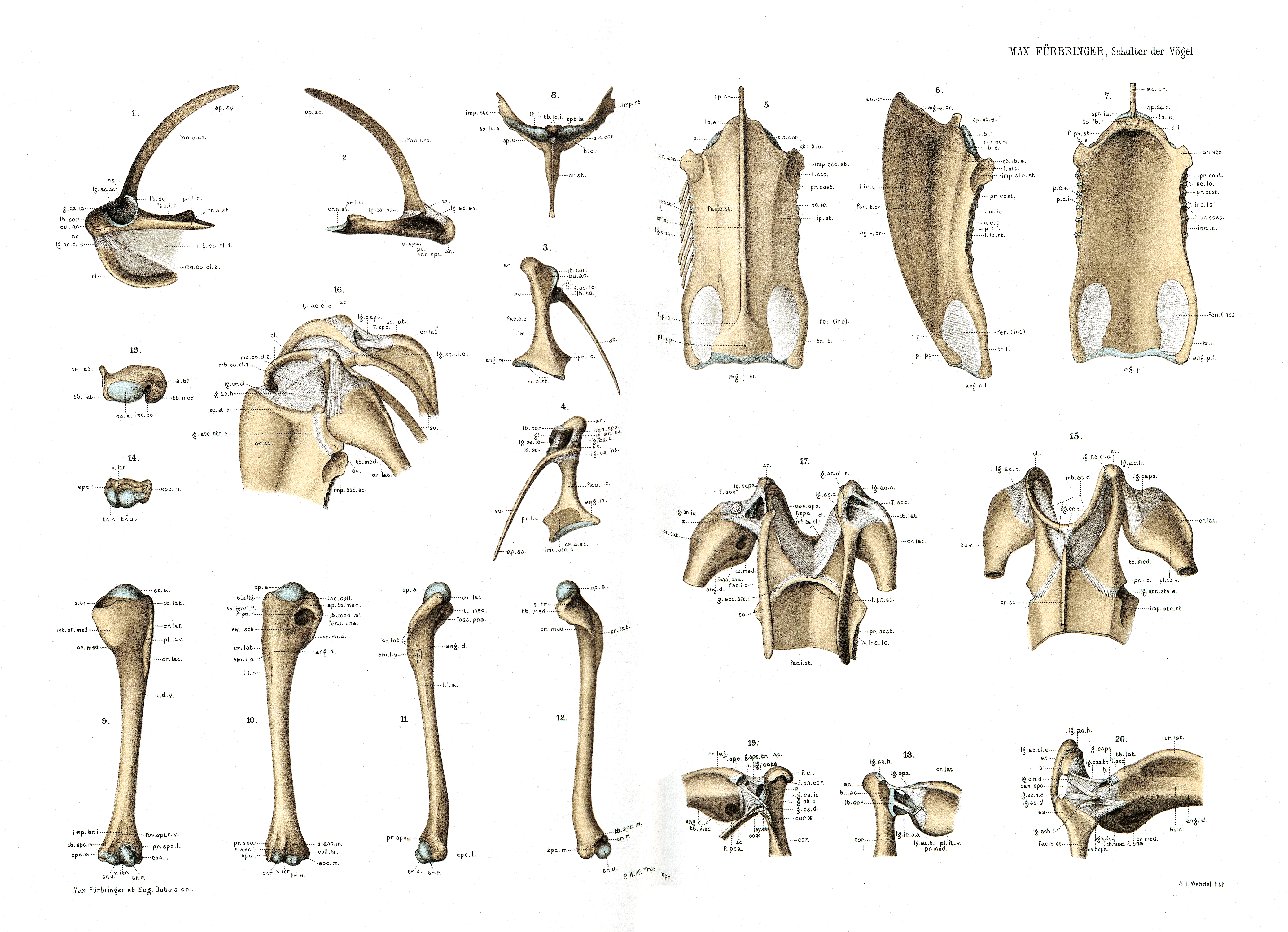
UNTERSUCHUNGEN ZUR MORPHOLOGIE UND SYSTEMATIK DER VÖGEL ac. Acrocoracoid (Carinaten) s. Spina coracoidea (Ratiten). ang. d. Angulus dorsalis des Humerus. ang. m. Angulus medialis distalis des Coracoid. ang. p. l. Angulus posterior lateralis des Sternum. ap. cr. Apex cristae sterni. ap. sc. Apex scapulae. as. Acromion. ap. th. med. Apex tuberculi medialis humeri. bu. ac. Stelle, wo die Bursa acrocoraeoidea dem Coracoid afliegt c., cor. Coracoid. can. spc. Canalis supracoraeoideus. cl. Clavicula. co., cor. Coracoid. coli. tr. Collum trochleae humeri. cost. Costae, Rippen. cp. a. Caput artieulare humeri er. a. st. Crista articularis sternalis des Coracoid Fig. 1. Coracoid, Scapula und Clavicula im Zusammenhange. Linke Seite; laterale Ansicht. 2. Coracoid und Scapula im Zusammenhange. Linke Seite; mediale Ansicht. 3. Coracoid und Scapula. Linke Seite; ventrale Ansicht. 4. Coracoid und Scapula. Links; dorsale (innere) Ansicht. 5. Sternum. Ventrale (äussere) Ansicht. 6. Sternum. Laterale Ansicht (von der linken Seite). 7. Sternum. Dorsale (innere) Ansicht. 8. Sternum. Ansicht von vorn (Gelenkfliichen für das Coracoid etc.). 9. Linker Humerus. Ventrale Ansicht. 10. Linker Humerus. Dorsale Ansicht. 11. Linker Humerus. Laterale Ansicht. 12. Linker Humerus. Mediale Ansicht. 13. Linker Humerus. Proximales Ende von oben. 14. Linker Humerus. Distales Ende von unten. 15. Brustgürtel, Brustbein und Humerus mit ihren Bandverbindungen. Ventrale (äussere) Ansicht. 16. Brustgürtel, Brustbein und Humerus mit ihren Bandverbindungen. Laterale Ansicht (von der linken Seite). 17. Brustgürtel, Brustbein und Humerus mit ihren Band Verbindungen. Dorsale (innere) Ansicht. 18. Schultergelenk (von Bernicla) von der ventralen Seite nach partieller Wegnahme des Lig. acrocoraco-humerale und der Kapsel. Humerus-Kopf etwas von der Fossa glenoidalis des Brustgürtels abgezogen. Linke Seite. 19. Schultergelenk (von Bernicla) von der dorsalen Seite nach Entfernung der Sehne des M. supracoracoideus und nach partieller Wegnahme des Acromion und des vorderen Endes des Coracoid. Humerus ebenfalls abgezogen. Linke Seite. 20. Schultergelenk (von Pandion) von der dorsalen Seite nach Wegnahme der Sehne des M. supracoracoideus. Humerus abgezogen. Rechte Seite. |
||||
| Author | Max Fuerbringer | ||||
| Permission (Reusing this file) |
|
||||
File history
Click on a date/time to view the file as it appeared at that time.
| Date/Time | Thumbnail | Dimensions | User | Comment | |
|---|---|---|---|---|---|
| current | 15:58, 23 January 2017 | | 4,158 × 3,009 (6.43 MB) | Shyamal (talk | contribs) | {{Information |Description=Shoulders of birds |Source=UNTERSUCHUNGEN ZUR MORPHOLOGIE UND SYSTEMATIK DER VÖGEL |Date=1888 |Author=Max Fuerbringer |Permission={{pd-old-70}} |other_versions= }} Category:Bird anatomy |
You cannot overwrite this file.
File usage on Commons
There are no pages that use this file.