File:Agarplate redbloodcells edit.jpg
Vai alla navigazione
Vai alla ricerca

Dimensioni di questa anteprima: 800 × 507 pixel. Altre risoluzioni: 320 × 203 pixel | 640 × 406 pixel | 1 024 × 649 pixel | 1 280 × 812 pixel | 2 000 × 1 268 pixel.
File originale (2 000 × 1 268 pixel, dimensione del file: 905 KB, tipo MIME: image/jpeg)
Didascalie
Didascalie
Aggiungi una brevissima spiegazione di ciò che questo file rappresenta
| Descrizione |
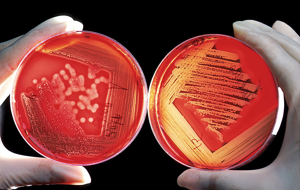
العربية: يُضاف دم الغَنم إلى المستنبت بَهدف تحسين العناصر الغذائية اللازمة للنمو البكتيري، حيثُ يَقوم علماء المُختبر بزرع وتنمية أنواع بكتيرية مُختلفة على هذا المستنبت وغيره من المستنبتات بغرض المُساعدة في تقييم العدوى البشرية والحيوانية. تُظهر الصُورة اليُمنى نمو البكتيريا العقدية حالة الدم-بيتا، حيثُ تقوم بإنتاج مُركب حال يعمل على تَحليل كريات الدم الحمراء المُحيطة بكل مُستعمرة. المستنبت مبقع كما ينبغي للحصول على مستعمرات بكتيرية معزولة. أما الصُورة اليُسرى تُظهر نمو أنواع المكورات العنقودية، فالمستنبت مُبقع بشكلٍ كثيف بالتالي لا يُمكن الحصول على مستعمرات بكتيرية معزولة.
English: Sheep blood is added to growth media to enhance the nutrients available for bacterial growth. Laboratory scientists grow various bacteria on this medium as well as numerous other media, in order to help assess infections in humans and animals. The image on the right shows growth of a beta-hemolytic streptococcus. The bacterium has produced a hemolytic compound that has lysed the red blood cells surrounding each colony. The plate was properly streaked to attain isolated colonies. The plate on the left is growing a Staphylococcus species. The plate was streaked too heavily, so isolated colonies were not attained. |
||||||
| Data | |||||||
| Fonte |
|
||||||
| Autore | Bill Branson – (Edited by Fir0002)(Edited by Drhx) | ||||||
| Licenza (Riusare questo file) |
|
||||||

|
Questa immagine è stata selezionata come Immagine del giorno in data 17 settembre 2009. La didascalia era la seguente: Italiano: Colture batteriche su due piastre di Petri ottenute da globuli rossi infetti. La piastra di sinistra mostra un'infezione da batteri del genere Staphylococcus, quella di destra da batteri del genere Streptococcus. Altre lingue:
Čeština: Krevní agar se používá k diagnostice infekcí. Petriho miska vlevo obsahuje stafylokokovou infekci, miska vpravo infekci streptokokem, charakteristické halo ukazuje na β-hemolytickou skupinu A. Dansk: Røde blodlegemer på en agarplade bruges til at diagnosticere infektioner. Pladen til venstre viser positiv staphylococcusinfektion, mens den højre viser positiv streptokokinfektion, hvor halo-effekten specifikt viser, at typen er beta-hæmolytisk gruppe A. English: Red blood cells on an agar plate are used to diagnose infection. The plate on the left shows a positive staphylococcus infection. The plate on the right shows a positive streptococcus infection and with the halo effect shows specifically a beta-hemolytic group A. Español: Glóbulos rojos (eritrocitos o hematíes) de la sangre en una placa de Petri con agar de las utilizadas para el diagnóstico de infecciones. La placa de la izquierda muestra una infección por estafilococos y la de la derecha, por estreptococos. Italiano: Colture batteriche su due piastre di Petri ottenute da globuli rossi infetti. La piastra di sinistra mostra un'infezione da batteri del genere Staphylococcus, quella di destra da batteri del genere Streptococcus. Magyar: Pozitív staphylococcus fertőzés (balra) és pozitív streptococcus fertőzés A-csoportú béta-hemolitikus udvarral (jobbra), véres agar táptalajon Nederlands: Rode bloedcellen op een agarplaat (petrischaal met een voedingsbodem zoals agaragar) voor het diagnosticeren van een infectie. De plaat aan linkerzijde toont een positieve stafylokokken-infectie. De plaat aan rechterzijde toont een positieve streptokokken-infectie, hetgeen door het halo-effect zichtbaar is als een beta-hemolytische uit de groep A (Streptococcus pyogenes). Deze infecties kunnen voorkomen bij patiënten die een chemokuur ondergaan. Polski: Czerwone krwinki na płytkach agarowych są używane do diagnozowania infekcji. Płytka po lewej wykazuje pozytywną infekcję gronkowcem (staphylococcus). Płytka po prawej wykazuje pozytywną infekcję paciorkowca (streptococcus) a efekt halo wskazuje na paciorkowce ropne (z grupy A). Português: Hemácias em uma placa de ágar são utilizadas para o diagnóstico de infecção. A placa da esquerda mostra uma infecção por estafilococos e a da direita, por estreptococos. Македонски: Црвените крвни зрнца на агарна плочка се користат за пронаоѓање на инфекции. На плочката лево е пронајден стафилокок, а на плочката десно се гледа присуство на стрептокок. Русский: Эритроциты используются для диагностики инфекций. Чашка Петри слева показывает наличие стафилококков, справа — стрептококков 한국어: 질병 감염을 진단하기 위해 사용되는 한천 판 위의 적혈구. 왼쪽 판은 포도상구균 감염 양성 반응을 보이고 있다. 오른쪽 판은 연쇄상구균 감염 양성 반응을 보이고 있고, 후광 효과를 통해 A군 베타용혈성 연쇄상구균임을 분명히 알 수 있다. (이 균은 성홍열을 일으킨다) فارسی : گویچههای سرخ در یک ظرف آگار برای تشخیص عفونت در یک آزمایشگاه مؤسسهٔ ملی سرطان آمریکا. ظرف سمت چپ محتوی عفونت مثبت استافیلوکک است و در ظرف سمت راست عفونت مثبت استرپتوکک دیده میشود. اینگونه عفونتها میتوانند در بیماران سرطانی، و بهصورت عوارض شیمیدرمانی ظاهر گردند. |
Cronologia del file
Fare clic su un gruppo data/ora per vedere il file come si presentava nel momento indicato.
| Data/Ora | Miniatura | Dimensioni | Utente | Commento | |
|---|---|---|---|---|---|
| attuale | 03:06, 6 gen 2008 | | 2 000 × 1 268 (905 KB) | Fir0002 (discussione | contributi) | == Summary == Public domain image from cancer.gov http://visualsonline.cancer.gov/details.cfm?imageid=2230. Red blood cells on an agar plate are used to diagnose infection. The plate on the left shows a positive staphyloccus infection. The plate on the r |
Impossibile sovrascrivere questo file.
Utilizzo del file
Le seguenti 36 pagine usano questo file:
- User talk:Durova/Archive 1
- Commons:Featured picture candidates/Image:Agarplate redbloodcells edit.jpg
- Commons:Featured picture candidates/Log/January 2008
- Commons:Featured pictures/Objects
- Commons:Featured pictures/chronological/2008-A
- Commons:Picture of the Year/2008/Galleries/All
- Commons:Picture of the Year/2008/Galleries/Objects
- Commons:Picture of the Year/2008/Results/Round 1/Gallery/All
- Commons:Picture of the Year/2008/Results/Round 1/Gallery/Objects
- Commons:Picture of the Year 2008/File:Agarplate redbloodcells edit.jpg
- Commons:Potd/2009-09 (da)
- Commons:Potd/2009-09 (de)
- Commons:Potd/2009-09 (eo)
- Commons:Potd/2009-09 (nl)
- Commons:Potd/2009-09 (pt)
- Commons:Potd/2009-09 (tr)
- File:Diagnostic algorithm of possible bacterial infection.png
- Template:Potd/2009-09
- Template:Potd/2009-09-17
- Template:Potd/2009-09-17 (cs)
- Template:Potd/2009-09-17 (da)
- Template:Potd/2009-09-17 (en)
- Template:Potd/2009-09-17 (es)
- Template:Potd/2009-09-17 (fa)
- Template:Potd/2009-09-17 (hu)
- Template:Potd/2009-09-17 (it)
- Template:Potd/2009-09-17 (ko)
- Template:Potd/2009-09-17 (mk)
- Template:Potd/2009-09-17 (nl)
- Template:Potd/2009-09-17 (pam)
- Template:Potd/2009-09-17 (pl)
- Template:Potd/2009-09-17 (pt)
- Template:Potd/2009-09-17 (ru)
- Template:Potd/2009-09-17 (zh-hans)
- Template:Potd/2009-09-17 (zh-hant)
- Template:Potd/2009-09 (zh-hans)
Utilizzo globale del file
Anche i seguenti wiki usano questo file:
- Usato nelle seguenti pagine di ar.wikipedia.org:
- بوابة:طب/صورة مختارة
- بوابة:علوم/صورة مختارة
- أغار
- مستنبت
- طبق آغار
- بوابة:علم الأحياء/صورة مختارة/أرشيف
- ويكيبيديا:صور مختارة/علوم/علم الأحياء
- بوابة:طب/صورة مختارة/6
- ويكيبيديا:ترشيحات الصور المختارة/كريات دم حمراء على طبق آجار
- ويكيبيديا:صورة اليوم المختارة/ديسمبر 2017
- قالب:صورة اليوم المختارة/2017-12-18
- بوابة:علوم/صورة مختارة/21
- بوابة:علم الأحياء/صورة مختارة/25
- ويكيبيديا:صورة اليوم المختارة/مارس 2020
- قالب:صورة اليوم المختارة/2020-03-05
- زراعة الأعشاب البحرية
- ويكيبيديا:صورة اليوم المختارة/يناير 2023
- قالب:صورة اليوم المختارة/2023-01-07
- قالب:صورة اليوم المختارة/تخطيط/2023/يناير
- Usato nelle seguenti pagine di bn.wikipedia.org:
- Usato nelle seguenti pagine di ca.wikipedia.org:
- Usato nelle seguenti pagine di crh.wikipedia.org:
- Usato nelle seguenti pagine di cs.wikipedia.org:
- Usato nelle seguenti pagine di cv.wikipedia.org:
- Usato nelle seguenti pagine di de.wikipedia.org:
- Usato nelle seguenti pagine di el.wiktionary.org:
- Usato nelle seguenti pagine di en.wikipedia.org:
- Agar
- Red
- Agar plate
- Portal:Medicine
- Growth medium
- Streaking (microbiology)
- Wikipedia:Featured pictures/Sciences/Biology
- Wikipedia:Featured pictures thumbs/09
- Wikipedia:Wikipedia Signpost/2008-01-14/Features and admins
- Wikipedia:WikiProject Media Restoration/Landmark images
- Wikipedia:Featured picture candidates/January-2008
- Wikipedia:Featured picture candidates/Image:Agarplate redbloodcells.jpg
- User talk:Durova/Archive 45
- User talk:Durova/Archive 51
- Wikipedia:Picture of the day/May 2008
- Template:POTD/2008-05-17
- User talk:Durova/Archive 57
- Wikipedia:Wikipedia Signpost/2008-01-14/SPV
Visualizza l'utilizzo globale di questo file.