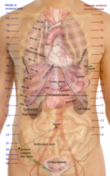
File:Surface projections of the organs of the trunk.svg

File originale (file in formato SVG, dimensioni nominali 475 × 760 pixel, dimensione del file: 3,2 MB)
Didascalie
Didascalie
Indice
Dettagli
[modifica]| DescrizioneSurface projections of the organs of the trunk.svg |
English: Surface projections of the major organs of the trunk, using the vertebral column and rib cage as main reference sources found in Superficial anatomy. The transpyloric plane and McBurney's point are among the marked locations.
For reference list and more information, see list on main page for the png-version To discuss image, please see Talk:Human body diagrams |
| Data | |
| Fonte | All used images are in public domain. |
| Autore | Mikael Häggström |
| Altre versioni |
  |
Licenza
[modifica]| Public domainPublic domainfalsefalse |
| Io, detentore del copyright su quest'opera, la rilascio nel pubblico dominio. Questa norma si applica in tutto il mondo. In alcuni paesi questo potrebbe non essere legalmente possibile. In tal caso: Garantisco a chiunque il diritto di utilizzare quest'opera per qualsiasi scopo, senza alcuna condizione, a meno che tali condizioni siano richieste dalla legge. |
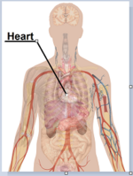
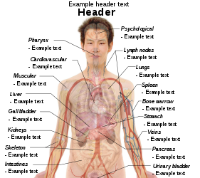
Human body diagrams[modifica]Main article at: Human body diagrams Template location:Template:Human body diagrams How to derive an image[modifica]Derive directly from raster image with organs[modifica]The raster (.png format) images below have most commonly used organs already included, and text and lines can be added in almost any graphics editor. This is the easiest method, but does not leave any room for customizing what organs are shown. Adding text and lines: Derive "from scratch"[modifica]By this method, body diagrams can be derived by pasting organs into one of the "plain" body images shown below. This method requires a graphics editor that can handle transparent images, in order to avoid white squares around the organs when pasting onto the body image. Pictures of organs are found on the project's main page. These were originally adapted to fit the male shadow/silhouette.
Organs:
Derive by vector template[modifica]The Vector templates below can be used to derive images with, for example, Inkscape. This is the method with the greatest potential. See Human body diagrams/Inkscape tutorial for a basic description in how to do this.
Examples of derived works[modifica]
Licensing[modifica]
|
Cronologia del file
Fare clic su un gruppo data/ora per vedere il file come si presentava nel momento indicato.
| Data/Ora | Miniatura | Dimensioni | Utente | Commento | |
|---|---|---|---|---|---|
| attuale | 09:19, 27 dic 2019 |  | 475 × 760 (3,2 MB) | Mikael Häggström (discussione | contributi) | +Costal margin |
| 10:38, 11 nov 2010 |  | 475 × 760 (3,2 MB) | Mikael Häggström (discussione | contributi) | Adapted to recently added overview images. Distinguished different ways to designate vertebrae levels. | |
| 10:04, 7 nov 2010 |  | 460 × 740 (3,16 MB) | Mikael Häggström (discussione | contributi) | heart in from of liver | |
| 09:41, 7 nov 2010 |  | 460 × 740 (3,16 MB) | Mikael Häggström (discussione | contributi) | Made heart a bit smaller. Marked sacral levels. | |
| 16:57, 4 nov 2010 |  | 460 × 740 (3,09 MB) | Mikael Häggström (discussione | contributi) | Lowered urinary bladder according to Fig 139 (see ref list) | |
| 04:28, 26 ott 2010 |  | 450 × 732 (3,04 MB) | Mikael Häggström (discussione | contributi) | symmetry | |
| 04:26, 26 ott 2010 |  | 450 × 732 (3,04 MB) | Mikael Häggström (discussione | contributi) | Moved T and L labels more to the right. Raised vertebral ending of some ribs to reach their correct level. Raised "stomach" label to avoid contact with the T10 line. | |
| 04:51, 24 ott 2010 |  | 517 × 732 (3,05 MB) | Mikael Häggström (discussione | contributi) | Smoother edges | |
| 05:18, 10 ott 2010 |  | 517 × 732 (3,02 MB) | Mikael Häggström (discussione | contributi) | Minor kidney adjustment. More realistic hip bone | |
| 05:35, 6 ott 2010 |  | 517 × 732 (2,77 MB) | Mikael Häggström (discussione | contributi) | Same scale as png-format |
Impossibile sovrascrivere questo file.
Utilizzo del file
Le seguenti 2 pagine usano questo file:
Utilizzo globale del file
Anche i seguenti wiki usano questo file:
- Usato nelle seguenti pagine di bew.wikipedia.org:
- Usato nelle seguenti pagine di en.wikipedia.org:
- Usato nelle seguenti pagine di es.wikipedia.org:
- Usato nelle seguenti pagine di incubator.wikimedia.org:
- Usato nelle seguenti pagine di uk.wikipedia.org:
Metadati
Questo file contiene informazioni aggiuntive, probabilmente aggiunte dalla fotocamera o dallo scanner usati per crearlo o digitalizzarlo. Se il file è stato modificato, alcuni dettagli potrebbero non corrispondere alla realtà.
| Larghezza | 475 |
|---|---|
| Altezza | 760 |


