Category:Cell biology
Naar navigatie springen
Naar zoeken springen

| Category Cell biology on sister projects: | |||||||||
|---|---|---|---|---|---|---|---|---|---|
Wiktionary |
Commons | ||||||||
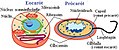
scientific discipline that studies cells  | |||||
| Media uploaden | |||||
| Uitspraak (audiobestand) | |||||
|---|---|---|---|---|---|
| Is een | |||||
| Subklasse van | |||||
| Onderdeel van | |||||
| Niet gelijk aan | |||||
| |||||
Ondercategorieën
Deze categorie bevat de volgende 118 subcategorieën, van de 118 in totaal.
*
A
- A549 cell line (2 B)
- Cell aggregation (103 B)
- Cell aging (17 B)
- Anisotropy in biology (8 B)
B
- Biological metamorphosis (23 B)
- Brochosome (6 B)
C
- Cell count (69 B)
- Cell degranulation (15 B)
- Cell disruption (17 B)
- Cell dynamics (5 B)
- Cell enlargement (19 B)
- Cell extracts (6 B)
- Cell fate determination (154 B)
- Cell fusion (109 B)
- Cell potency (52 B)
- Cell tracking (43 B)
- Cellular immunity (8 B)
- Cellular stress responses (247 B)
- Chemiosmosis (18 B)
- Colony-forming units assays (21 B)
- Cytolysis (3 B)
D
E
- Endomembrane system (10 B)
- Extracellular traps (22 B)
F
G
- Goodsell molecular landscape (14 B)
H
I
- Intracellular space (82 B)
M
- Media from BMC Cell Biology (220 B)
- Media from Cell & Bioscience (4 B)
- Media from Cell & Chromosome (4 B)
- Media from Cell Regeneration (2 B)
O
- Oogenesis (42 B)
- Osmotic shock (6 B)
P
- Cell-penetrating peptides (12 B)
- Phytoliths (14 B)
- Plasmolysis (24 B)
- Prebiotic (316 B)
- Protoplasts (18 B)
R
S
- Cell shape (259 B)
- Single-cell analysis (80 B)
- Subcellular fractions (57 B)
- Subcellular localization (10 B)
- Surface properties (biology) (7 B)
- Cell survival (228 B)
T
V
- Videos of cell biomechanics (13 B)
Z
- ZTZ der Uniklinik Freiburg (6 B)
Pagina’s in categorie "Cell biology"
Deze categorie bevat alleen de volgende pagina.
Media in categorie "Cell biology"
Deze categorie bevat de volgende 200 bestanden, van in totaal 545.
(vorige pagina) (volgende pagina)-
 022S.jpg 1.772 × 1.772; 3,11 MB
-
 023S.jpg 1.772 × 1.792; 612 kB
-
 1 - ID applications.png 1.662 × 950; 459 kB
-
 200801large.jpg 600 × 457; 298 kB
-

-
 208031 EPFL David Suter Sox2.jpg 1.304 × 734; 52 kB
-
 3eme vague.png 550 × 357; 7 kB
-
 41598 2018 20107 Fig3 HTML.jpg 660 × 315; 167 kB
-
 46085 orig.jpg 2.816 × 2.112; 412 kB
-
 72hr sodyum48 saat.jpg 1.600 × 1.200; 272 kB
-
 A Critical-like Collective State Leads to Long-range Cell Communication in Dictyostelium discoideum Aggregation.pdf 1.275 × 1.650, 19 pagina's; 3,34 MB
-
 A sol mit Paramecium.jpg 1.327 × 885; 241 kB
-
 A) Endocitosis y b) exocitosis.jpg 818 × 529; 109 kB
-
-
 A549 bridges -- 7-28-at1622.jpg 1.392 × 1.040; 160 kB
-
 A549 bridges -- 7-28-at1623.jpg 1.392 × 1.040; 149 kB
-

-
 Adrenergic signaling on natriuretic peptides.jpg 2.958 × 2.165; 711 kB
-
 Alcatel One Touch 720.pdf 1.239 × 1.752, 4 pagina's; 48 kB
-
 Allosteric regulation mode, feedback inhibition and its reversal.png 709 × 499; 72 kB
-
 ALPSc.jpg 373 × 197; 19 kB
-
 Alveolar sac region of the lung - TEM.jpg 1.560 × 1.257; 522 kB
-
 Amiloplastos de células de papa.jpg 302 × 325; 53 kB
-
 Animal Cell Structure.png 724 × 484; 145 kB
-
 Annotated structure of eRF1.jpg 600 × 361; 81 kB
-
 Annular Gap Junction Vesicle.jpg 205 × 153; 61 kB
-
 Anthracologie-exemple.jpg 860 × 860; 848 kB
-
 Anthracologie-exemple2.jpg 896 × 896; 786 kB
-
 Anticossos anticardiolipina.jpg 427 × 286; 61 kB
-
 Apical Constriction.jpg 242 × 300; 80 kB
-
 Apicalconstriction fig1.jpg 417 × 467; 82 kB
-
 Apicalconstriction fig2.jpg 393 × 437; 82 kB
-
 Apoptotic DNA Laddering.png 178 × 193; 20 kB
-
 ART SCIENCE Craig Hilton 'The Immortalisation of Billy Apple' 01.jpg 3.264 × 2.448; 2,91 MB
-
 ART SCIENCE Craig Hilton 'The Immortalisation of Billy Apple' 02.jpg 3.264 × 2.448; 3,01 MB
-
-
 Aurora B localization.jpg 93 × 349; 8 kB
-
 Autophagy and apoptosis.png 678 × 339; 97 kB
-
 Autophagy and cancer.jpg 985 × 380; 76 kB
-
 Autophagy in plants.jpg 600 × 424; 53 kB
-
 Autophagy's function.gif 200 × 98; 11 kB
-
 Autophagy.png 2.012 × 1.636; 1,59 MB
-
 Axopodium Mikrotubuli.jpg 748 × 682; 269 kB
-
 Banner cell biology.png 1.003 × 100; 45 kB
-
 BarrBodyBMC Biology2-21-Fig1.jpg 1.200 × 1.417; 298 kB
-
 Betareg.PNG 523 × 511; 18 kB
-
 BiggeggSH-SY5Y.jpg 1.600 × 1.200; 426 kB
-
 BioArchive.jpg 263 × 342; 31 kB
-
 Blood cell crossing vascular sinus wall - TEM.jpg 1.560 × 1.254; 518 kB
-
 Boveri-signature.jpg 424 × 91; 20 kB
-
 Branching morphogenesis in 3D cell culture.jpg 778 × 434; 98 kB
-
 Brefeldin A Inhibition of Intracellular Vesicle Transport.png 591 × 271; 23 kB
-
 Brochosome model1.jpg 729 × 360; 40 kB
-
 BrUpolIIc.jpg 406 × 233; 33 kB
-
 BS-Fig1.jpg 658 × 336; 105 kB
-
 Cajal frontal lobe.gif 250 × 442; 39 kB
-
 Cajal Purkinje.gif 300 × 332; 32 kB
-
 Calcium Signaling Pathway.png 1.526 × 907; 180 kB
-
 Cancer type vs frequency.png 943 × 354; 25 kB
-
 Catenin-humanendothel.jpg 800 × 634; 376 kB
-
 CBDS.Mirmira-300x167.png 300 × 167; 58 kB
-
 Cel cible compétente.png 854 × 165; 6 kB
-
 Celcultuuroven.JPG 2.560 × 1.920; 1,94 MB
-
 Celkweekverversing.jpg 1.920 × 2.560; 279 kB
-
 Cell (PSF).jpg 969 × 430; 386 kB
-
 Cell 1 a.jpg 373 × 200; 46 kB
-
 Cell 1.jpg 1.500 × 805; 654 kB
-
 Cell structure.jpg 1.280 × 720; 127 kB
-
 Cell-organelles-labeled.png 1.050 × 1.024; 743 kB
-
 Cell-organelles.webp 600 × 375; 15 kB
-
 Cell-shape-mitosis.png 633 × 668; 167 kB
-
 Cell-type specificity of TIP-YFP expression in the root axis cropped.jpg 1.050 × 565; 120 kB
-
 Cell-type specificity of TIP-YFP expression in the root axis.jpg 1.200 × 2.007; 554 kB
-
 Cell-universe.jpg 1.398 × 1.045; 51 kB
-
 Cells detention centers 2.jpg 527 × 265; 87 kB
-
 Cells detention centers 3.jpg 300 × 151; 65 kB
-
 Cells detention centers.jpg 344 × 172; 59 kB
-
 Cells in space.JPG 1.280 × 960; 398 kB
-
 Cellsize it.jpg 310 × 199; 10 kB
-
 Cellsize PL.png 310 × 199; 72 kB
-
 Cellsize.jpg 310 × 199; 51 kB
-
 Celltype zh.png 1.280 × 535; 176 kB
-
 Celltypes rus.png 468 × 202; 51 kB
-
 Cellular Dewetting.jpg 606 × 102; 14 kB
-
 Cellular Structure (2).jpg 5.312 × 2.988; 4,08 MB
-
 Cellular Structure.jpg 5.243 × 2.854; 3,11 MB
-
 Cellular tight junction-cz.svg 499 × 646; 145 kB
-
 CELLULES ROUGES.jpg 785 × 785; 466 kB
-
 Cellules souches embryonnaires HD90.jpg 2.048 × 1.536; 623 kB
-
 Cellules souches embryonnaires HD90.tif 2.048 × 1.536; 6 MB
-
 Celulasok.jpg 2.491 × 1.072; 3,59 MB
-
 Chaperiony.png 794 × 1.123; 77 kB
-
 CHIB.-Sander-300x226.jpg 300 × 226; 25 kB
-
 CHIB.-Stabler.jpg 1.362 × 627; 675 kB
-
 Chloride cell.jpg 574 × 480; 276 kB
-
 Cho cells adherend1.jpg 1.280 × 960; 387 kB
-
 Cho cells adherend2.jpg 1.280 × 960; 240 kB
-
 Chromaffin cell imaged with DIC and IRM.png 330 × 212; 63 kB
-
 Cineálacha éagsúla ceall.jpg 450 × 188; 43 kB
-
 Città della Scienza Catania 2.jpg 2.896 × 1.944; 1,49 MB
-
 Cleavage-furrow.JPG 449 × 447; 23 kB
-
 Clonal expansion and monoclonal versus polyclonal proliferation.PNG 660 × 510; 34 kB
-
 Cluster of Solenocytes.jpg 862 × 930; 152 kB
-
 CnGRASP55domainsc.jpg 763 × 473; 112 kB
-
 CollagenMegaCarrierc.jpg 553 × 249; 45 kB
-
 Colonies of Madin-Darby Canine Kidney cells grown in tissue culture.jpg 1.030 × 1.030; 263 kB
-
 Color fungi.jpg 1.503 × 1.127; 452 kB
-
 Comparison chrx.jpg 1.114 × 729; 491 kB
-

-
 Complete Hydatidiform Mole (38711526030).jpg 1.732 × 2.048; 1,34 MB
-
 Conger type callus 3ms White Light.TIF 2.048 × 1.536; 9,01 MB
-
 Conidiospore-hyaloperonospora-parasitica-appressorium.jpg 590 × 233; 53 kB
-
 Cotyledon-Cercis siliquastrum.jpg 772 × 516; 243 kB
-
 Coxiella burnetii, the bacteria that causes Q Fever.jpg 2.424 × 2.032; 1,19 MB
-
 CPE rounding.jpg 1.200 × 900; 366 kB
-
 CPE syncytium.jpg 1.200 × 900; 468 kB
-
 Cresta Localiza ATPsintasa.png 580 × 440; 126 kB
-
 Cresta Mitocondrial.png 1.123 × 794; 293 kB
-
 Crosssectionroottooth11-24-05.jpg 1.263 × 1.053; 259 kB
-
 Crosstalk TM.png 1.588 × 616; 182 kB
-
 Cryptosporidium parvum auramine-rhodamine labeled.jpg 300 × 308; 6 kB
-
 CTAR Powers.png 1.173 × 639; 1,19 MB
-
 CTAR.-Herrera-300x165.png 300 × 165; 50 kB
-
 Cyborg Cell characteristics.jpg 501 × 510; 143 kB
-
 Cyto.png 2.426 × 577; 98 kB
-
 Cytogenetic Telomere Arrays (2005).jpg 990 × 1.333; 200 kB
-
 Cytokinesis-electron-micrograph.jpg 745 × 451; 200 kB
-
 Cytoneme.tif 1.400 × 796; 1,28 MB
-
 Células en un tejido normal.jpg 1.280 × 720; 39 kB
-
 Dedication to Plant Cell Biology Second Edition, Elsevier 2019.jpg 4.383 × 6.284; 3,32 MB
-
 Dental Pulp cultured by MLM (3D vs. 2D) - 5 Days.tiff 1.353 × 1.112; 1,18 MB
-
 Desmosome cell junction cs.svg 556 × 588; 124 kB
-
 Desplazamiento del punto central de la célula.gif 724 × 427; 11 kB
-

-

-
 Different ways mtDNA moves into the nucleus.PNG 1.280 × 720; 89 kB
-
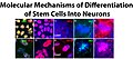 Differentiation of Stem Cells Into Neurons.jpg 1.884 × 835; 845 kB
-
 Differentiation.jpg 5.454 × 3.225; 31,2 MB
-
 DnTRFc.jpg 462 × 259; 34 kB
-
 Domain architecture and structure of C-terminal EHD proteins.gif 583 × 522; 41 kB
-
 Drachenbaum Bewurzelung-in-Wasser Kallus SL273382.JPG 2.304 × 3.072; 2,23 MB
-
 Drawing of nucleus.jpg 2.448 × 1.836; 1,05 MB
-
 Duplicazione dei plasmidi.png 800 × 700; 223 kB
-
 Débit de Filtration Glomérulaire Inkscape.png 2.500 × 1.500; 544 kB
-
 Débit de Filtration Glomérulaire librsvg.png 2.500 × 1.500; 458 kB
-
 Débit de Filtration Glomérulaire rendersvg.png 2.500 × 1.500; 476 kB
-
 EHD proposed mechanism.jpg 1.280 × 373; 70 kB
-
 Einzeller 800 fach Streiflicht.jpg 1.952 × 1.372; 409 kB
-
 Ekstratsellulaarne maatriks.png 800 × 554; 97 kB
-
 Electron micrograph of a Fractone.jpg 3.662 × 3.472; 2,37 MB
-
 Embolized Uterus (44319080955).jpg 1.270 × 1.124; 808 kB
-
 Embolized Uterus (44319081065).jpg 1.352 × 1.124; 811 kB
-
 Embolized Uterus (44507244194).jpg 1.124 × 1.208; 885 kB
-
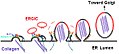
 EndoERGICNIHlc.jpg 273 × 291; 23 kB
-
 Endometrial Intraepithelial Neoplasia (EIN) (46262991251).jpg 2.347 × 1.706; 1,61 MB
-
 Endosymbiose.jpg 1.899 × 285; 198 kB
-
 Entropy in living cells.jpg 897 × 606; 93 kB
-
 Eosonophilic promyelocyte.png 115 × 104; 12 kB
-
 Epitelo-mezenchymální tranzice.gif 822 × 214; 36 kB
-
 Epithelium TCJ.png 755 × 736; 969 kB
-
 Epithelzelle 43729.jpg 531 × 384; 24 kB
-
 Epithelzellen16631.jpg 531 × 384; 33 kB
-
 ER-containing autophagosome.png 1.417 × 1.770; 2,06 MB
-
 ERK propagation waves from the wound edge.png 1.340 × 547; 1,07 MB
-
 Ethanol-Bilayer.png 1.194 × 639; 265 kB
-

-
 Farlik abstract.jpg 375 × 375; 71 kB
-
 FGF Pathway tm.png 1.242 × 698; 58 kB
-
 Fibers of Collagen Type I - TEM .jpg 640 × 480; 105 kB
-
 Fibers of Collagen Type I - TEM.jpg 640 × 480; 100 kB
-
 Fibroblast-2.jpg 2.999 × 2.249; 693 kB
-
 Flow cytometer structure.png 865 × 599; 1,98 MB
-
 Flowcytometer MoFlo (DAKO cytomation).jpg 320 × 426; 21 kB
-
 Flower petal cells image 2.jpg 3.072 × 4.096; 5,6 MB
-
 Flower petal cells.jpg 3.072 × 4.096; 4,75 MB
-
 Fluorescence.microscope1.jpg 2.226 × 2.544; 1,09 MB
-
 Fluorescence.microscope2.jpg 2.428 × 2.475; 1,21 MB
-
 Fluorescence.microscope3.jpg 2.436 × 2.393; 1,26 MB
-
 Food particle from a colonoscopy specimen (34016474661).jpg 1.480 × 811; 369 kB
-
 Foreign Body Granuloma on the Peritoneum (41962302110).jpg 2.402 × 1.458; 1,28 MB
-
 Formacao Mesossomo.png 900 × 800; 63 kB
-
 Formation of the new blood vessels.jpg 1.920 × 1.200; 2,81 MB
-
 FTIR of cell.png 3.025 × 2.266; 2,28 MB
-
 Functions of DS.PNG 585 × 402; 104 kB
-
 Fusion secuencia.jpg 2.084 × 1.828; 2,62 MB
-
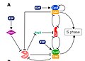 G1-S cell cycle regulation.jpg 800 × 576; 68 kB
-
 G2-M Bistability.png 1.347 × 1.012; 49 kB
-
 Ganglion Cyst of Hand (35295313226).jpg 2.102 × 1.669; 659 kB
-
 Gauchdisease.jpg 314 × 209; 74 kB
-
 GemIdentComposite.jpg 744 × 683; 125 kB
-
 Gephyrinc.jpg 542 × 347; 52 kB
-
 Gewebetypen des Blattes.jpg 3.508 × 2.480; 1,28 MB
-
 Glycophosphatidylinositol anchor.tif 585 × 432, 2 pagina's; 514 kB
-
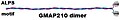
 GMAP210ALPSc.jpg 376 × 188; 19 kB
-
 GMAP210c.jpg 420 × 71; 11 kB
-
 Gnf-segmented-41-closeup.png 351 × 236; 83 kB
-
 GolgiCisConnc.jpg 330 × 123; 21 kB
-
 GolgiCOGsc.jpg 748 × 324; 84 kB
-
 GolgiRibbonc.jpg 250 × 143; 23 kB
-
 GolgiScyl1c.jpg 471 × 296; 46 kB


_Endocitosis_y_b)_exocitosis.jpg)


.jpg)


.jpg)


.jpg)


.jpg)


.jpg)
.jpg)
.jpg)

_(46262991251).jpg)


.jpg)


.jpg)
.jpg)


.jpg)


