Category:Embryos
Jump to navigation
Jump to search
multicellular diploid eukaryote in its earliest stage of development  | |||||
| Upload media | |||||
| Instance of |
| ||||
|---|---|---|---|---|---|
| Subclass of |
| ||||
| Location of creation | |||||
| Follows | |||||
| Followed by |
| ||||
| Different from | |||||
| Said to be the same as | Q12887356 | ||||
| |||||
Subcategories
This category has the following 10 subcategories, out of 10 total.
Media in category "Embryos"
The following 105 files are in this category, out of 105 total.
-
 2-Cell Love.jpg 955 × 955; 170 KB
-
 4-cell stage embryo.png 500 × 375; 87 KB
-
 4cell embryo.tif 1,199 × 1,225; 4.2 MB
-
 6 week embryo brain-ar.jpg 544 × 401; 44 KB
-
 8-cell stage embryo.png 500 × 375; 94 KB
-
 Acquapendente de formatu2.png 374 × 575; 202 KB
-
 Agelena labyrinthica embryo (01).jpg 822 × 688; 559 KB
-
 Agelena labyrinthica embryo.jpg 833 × 647; 688 KB
-
 Agelena labyrinthica four stages in the development.jpg 988 × 691; 718 KB
-
 Agelena labyrinthica procephalic lobes embryo.jpg 1,143 × 500; 638 KB
-
 Agelena labyrinthica thoracic region embryo.jpg 882 × 717; 512 KB
-
 Agelena labyrinthica twor stages in the laate development.jpg 863 × 645; 464 KB
-
 Agelena labyrinthica ventral plate at three stages.jpg 1,120 × 719; 454 KB
-
 Amniotic sac cow.jpg 5,568 × 3,712; 3.54 MB
-
 Anodonta piscinalis segmentation embryo.jpg 1,287 × 379; 435 KB
-
 Astacus development embryo.jpg 984 × 740; 349 KB
-
 Astacus embryo.jpg 543 × 818; 399 KB
-
 Atlas de Embrião de Camundongo E14.5.png 850 × 444; 147 KB
-
 Brinster Mouse Egg Culture 1963 slides.png 1,060 × 1,125; 1.77 MB
-
 Bunga kelapa.jpg 2,250 × 4,000; 1.38 MB
-
 Calopteryx embryo development.jpg 669 × 740; 429 KB
-
 Cat Diagram of foetus, showing the visceral arches and budding limbs.jpg 688 × 741; 475 KB
-
 Cat longitudinal section through the axis of the ovum.jpg 878 × 706; 839 KB
-
 Cat medullary groove.jpg 1,550 × 640; 449 KB
-
 Chelifer embryo.jpg 967 × 793; 820 KB
-
 Cnidariangastrula.jpg 400 × 400; 52 KB
-
 Comparative somite creation.jpg 775 × 447; 78 KB
-
 Cucumaria doliolum embryo at the end of the fourth day.jpg 604 × 689; 513 KB
-
De-Embryo.ogg 1.5 s; 13 KB
-
De-Leibesfrucht.ogg 2.2 s; 21 KB
-
 Descent fig02.jpg 344 × 702; 74 KB
-
-
 Diseases of the horse's foot (Page 54) BHL23615248.jpg 2,307 × 3,676; 876 KB
-
Drosophila cleavage and gastrulation.webm 30 s, 1,920 × 800; 42.28 MB
-
 E14.5.png 850 × 524; 314 KB
-
 Echinoderm embryo undergoing second cleavage.jpg 1,740 × 1,700; 532 KB
-
 Eight- Cell Embryo-Blastomeres.jpg 376 × 376; 21 KB
-
 Elements of the comparative anatomy of vertebrates (1886) (21057027940).jpg 1,328 × 1,500; 342 KB
-
 Embrione di vitello di circa 1 mese (diametro 22 millimetri) nel sacchetto amniotico.jpg 4,288 × 3,216; 3.81 MB
-
-
 Embrião de camundongo E14.5 em desenvolvimento.jpg 221 × 344; 6 KB
-
 Embrião de Rã - Secção da Cabeça.png 776 × 520; 838 KB
-
 Embrião de Rã - Secção do Abdómen.png 639 × 805; 1.12 MB
-
 Embrião de Rã - Secção do Tórax.png 560 × 790; 767 KB
-
 Embryo - copies.jpg 2,652 × 2,904; 2.61 MB
-
 Fish market in a road.jpg 720 × 707; 143 KB
-
 Gebärmutter mit Embryo im Längsschnitt - Uterus longitudinal section with embryo.jpg 1,961 × 1,735; 525 KB
-
 Gray26.svg 344 × 345; 21 KB
-
 Gray27.png 300 × 298; 10 KB
-
 Gray28.svg 396 × 455; 14 KB
-
 Haeckel Thoracostraca.jpg 2,342 × 3,289; 1.54 MB
-
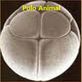 Holobastica polo animal.png 143 × 143; 45 KB
-
 Holothuria tubulosa development embryo two stages.jpg 1,079 × 688; 735 KB
-
 Holothuria tubulosa development embryo.jpg 1,224 × 675; 776 KB
-
 Human blastoid - 1.jpg 2,835 × 2,835; 2.42 MB
-
 Human blastoid.jpg 1,089 × 1,089; 1.24 MB
-
 Hydrophilus piceus embryos (01).jpg 754 × 750; 520 KB
-
 Hydrophilus piceus embryos.jpg 815 × 657; 616 KB
-
 Images representing technical steps during sEmbryo culture protocol.jpg 3,206 × 4,151; 1.98 MB
-
 Loligo advanced embryo.jpg 515 × 862; 500 KB
-
-
 Many human blastoids.jpg 1,500 × 671; 620 KB
-
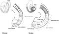
 Mouse and Snake Embryos.jpg 867 × 706; 49 KB
-
 Mouse embryo vasculature.tif 788 × 975; 1.1 MB
-
 Má-formações Nêurula.png 355 × 434; 8 KB
-

-
 Neural crest.png 450 × 540; 39 KB
-
 Neurula.png 873 × 317; 21 KB
-
 Oniscus murarius embryo.jpg 1,488 × 667; 1.13 MB
-
 P. miniata journey to metamorphosis - 2 days.tif 1,920 × 1,080; 5.94 MB
-
 P. miniata journey to metamorphosis - 4 days.tif 1,920 × 1,080; 5.94 MB
-
 Palaemon development embryo (01).jpg 1,356 × 679; 1,001 KB
-
 Palaemon development embryo.jpg 1,279 × 662; 828 KB
-
 Paracentrotus lividus cleavage stadia.jpg 1,282 × 870; 1.05 MB
-
 Paracentrotus lividus life cycle.jpg 725 × 693; 204 KB
-
 Perdeyên embriyonî û plasenta ku.png 1,003 × 498; 395 KB
-
 Plate IV. Diagrammatic transverse sections of different embryos.jpg 1,071 × 1,648; 1.32 MB
-
 Plate V. Diagrammatic longitudinal sections of different embryos.jpg 1,389 × 2,174; 1.62 MB
-

-
 PSM V71 D367 Three sections through a rabbit embryo of seven and half days.png 1,673 × 1,647; 455 KB
-
 PSM V71 D465 Nuclei from rabbit embryos.png 1,654 × 1,590; 303 KB
-
 Rana temporaria cleavage at the close of segmentation embryo.jpg 768 × 727; 627 KB
-
 Rana temporaria cleavage embryo.jpg 1,361 × 603; 800 KB
-
-
 Schrader; embryology Wellcome L0000395.jpg 1,142 × 1,814; 819 KB
-
 Scorpion embryo enveloped in its membranes.jpg 627 × 787; 683 KB
-
 Scorpion embryo mesoblastic somites.jpg 527 × 736; 354 KB
-
 Scorpion embryo.jpg 936 × 715; 828 KB
-
 Scorpion ovum with blastoderm.jpg 553 × 675; 427 KB
-
 Scorpion ventral plate.jpg 624 × 653; 361 KB
-
 Sea urchin embryo.jpg 1,430 × 1,096; 703 KB
-
 Seeds of orchids (J.G.Beer -1863).jpg 600 × 900; 78 KB
-
 Strongylosoma guerinii development embryo (01).jpg 1,120 × 706; 803 KB
-
 Strongylosoma guerinii development embryo.jpg 1,077 × 527; 674 KB
-
 The American journal of anatomy (1909) (17534170593).jpg 1,768 × 3,016; 844 KB
-

-
 The pedigree of man - and other essays (1903) (14578076870).jpg 2,448 × 1,342; 792 KB
-
 Tornaria early stage in the development embryo (01).jpg 660 × 687; 427 KB
-
 Tornaria early stage in the development embryo.jpg 618 × 667; 429 KB
-
 Tornaria late stage in the development embryo.jpg 477 × 793; 446 KB
-
 Tornaria stages in the development embryo.jpg 658 × 751; 381 KB
-
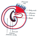
 Vetebrate Embryo.jpg 960 × 720; 72 KB
-
 Vetebrateembryo.svg 716 × 604; 197 KB
-
 Çeqîn ku.png 594 × 653; 133 KB
-
 Эмбрион позвоночных (изм.).png 3,000 × 2,250; 2.57 MB


.jpg)


_BHL23615248.jpg)


_(21057027940).jpg)
_nel_sacchetto_amniotico.jpg)
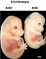
_e_deficiente_de_Ant2%2B.jpg)


.jpg)


.jpg)


.jpg)
.jpg)

_(17534170593).jpg)
_BHL16226079.jpg)
_(14578076870).jpg)
.jpg)


.png)