File:Fundus photograph of a healthy right eye (OD) from a myopic female Asian patient. Age 22.png
Jump to navigation
Jump to search
Size of this preview: 800 × 531 pixels. Other resolutions: 320 × 213 pixels | 640 × 425 pixels | 1,024 × 680 pixels | 1,280 × 850 pixels | 2,560 × 1,700 pixels | 4,288 × 2,848 pixels.
Original file (4,288 × 2,848 pixels, file size: 7.47 MB, MIME type: image/png)
Captions
Captions
Add a one-line explanation of what this file represents
Summary[edit]
_from_a_myopic_female_Asian_patient._Age_22.png&action=edit§ion=1)
| Description |
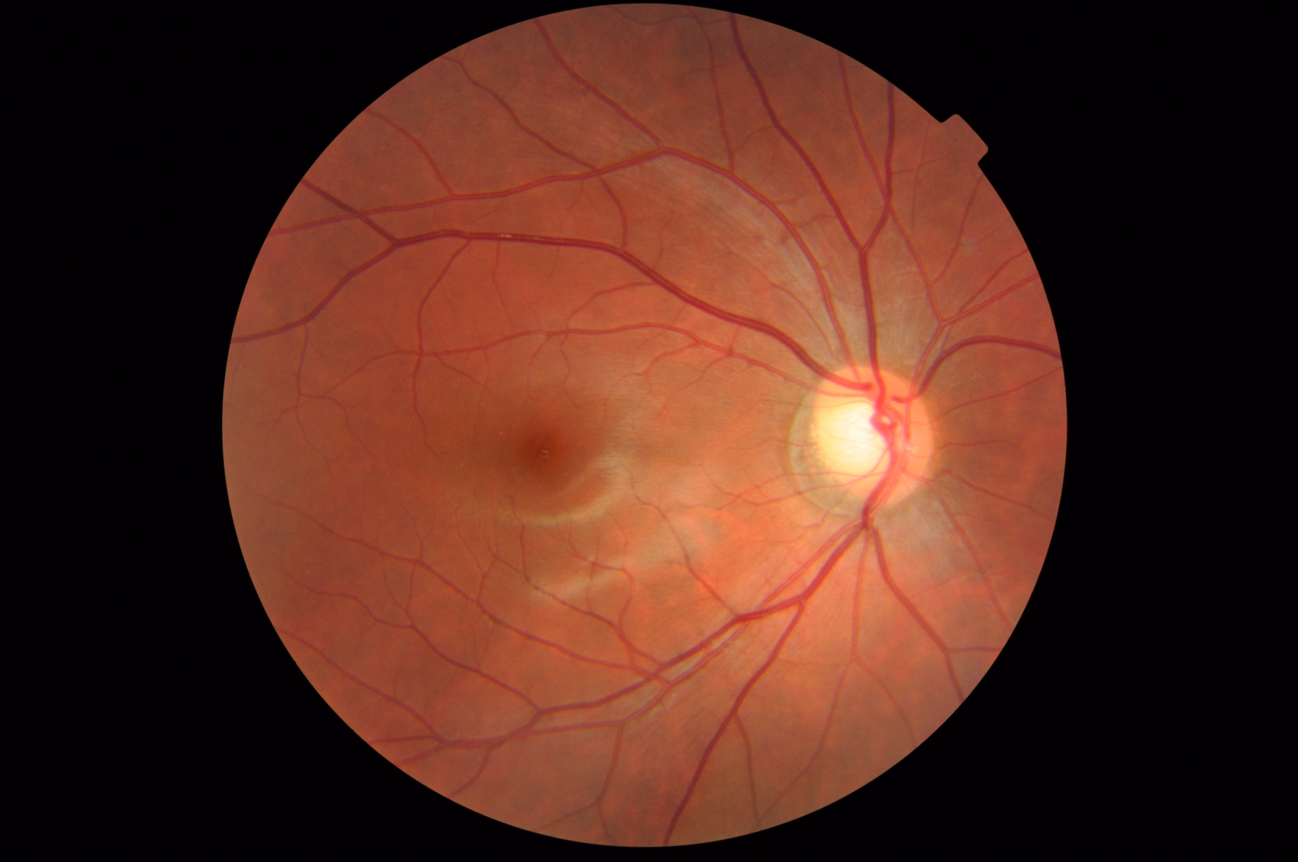
Fundus photograph of a healthy right eye (OD) from a myopic female Asian patient. Age 22. The white glial tissue indicates a healthy nerve fiber layer above the retinal blood vessels. The large cup within the disc is sloping temporally. The macular region is clear and has a visible fovea. Lamina is visible. Circumlinear vessels along the superior and inferior edges of the cup. Keywords: fundus, photograph, healthy, glial, posterior, pole, back, eye, retina, optic disc, macula, lamina, circumlinear vessels |
| Date | |
| Source | Fundus Photo, Right Eye (OD) |
| Author | OptometrusPrime |
Licensing[edit]
_from_a_myopic_female_Asian_patient._Age_22.png&action=edit§ion=2)
This file is licensed under the Creative Commons Attribution-Share Alike 2.0 Generic license.
- You are free:
- to share – to copy, distribute and transmit the work
- to remix – to adapt the work
- Under the following conditions:
- attribution – You must give appropriate credit, provide a link to the license, and indicate if changes were made. You may do so in any reasonable manner, but not in any way that suggests the licensor endorses you or your use.
- share alike – If you remix, transform, or build upon the material, you must distribute your contributions under the same or compatible license as the original.
| This image was originally posted to Flickr by OptometrusPrime at https://www.flickr.com/photos/85105636@N07/7805918268. It was reviewed on 27 January 2013 by FlickreviewR and was confirmed to be licensed under the terms of the cc-by-sa-2.0. |
File history
Click on a date/time to view the file as it appeared at that time.
| Date/Time | Thumbnail | Dimensions | User | Comment | |
|---|---|---|---|---|---|
| current | 10:06, 27 January 2013 | | 4,288 × 2,848 (7.47 MB) | Jacopo Werther (talk | contribs) | {{Information |Description= Fundus photograph of a healthy right eye (OD) from a myopic female Asian patient. Age 22. The white glial tissue indicates a healthy nerve fiber layer above the retinal blood vessels. The large cup within the disc is sloping... |
You cannot overwrite this file.
File usage on Commons
The following page uses this file:
_from_a_myopic_female_Asian_patient._Age_22.png&oldid=760879781)